Symptoms
In Achalasia, you would find it difficult to swallow; it feels like the food is stuck in your food pipe. This condition is referred to as ‘Dysphagia.' This happens because of the reduced movement in the Oesophagus (peristalsis). Due to Dysphagia, there can be an increased risk of aspiration (inhalation of Gastric contents).
The common symptoms include:
- Cough
- Hiccups
- Chest pain
- Heartburn
- Weight loss
- Sensation of fullness
- Difficulty in swallowing
- Regurgitation (reverse flow) of food
Causes
The cause of Achalasia is unknown. So, it could be difficult for your doctor to find out the specific cause. The most common causes include:
- Heredity
- Chagas’ disease
- Autoimmune condition
- Damage to Oesophageal nerves
- Lack of normal contractions in Oesophageal muscles
Risk factors
The most common risk factor for Achalasia is the presence of Autoimmune Disorders.
Achalasia mostly occurs in your middle to old age i.e., 25 to 60 years. It can also occur in children although.
Complications
Some of the most common complications of Achalasia include:
- Weight loss
- Malnutrition
- Oesophagitis
- Breathlessness
- Pulmonary infection
- Aspiration Pneumonia
Medical help
Call your health care provider if:
- You have difficulty during swallowing
- Your symptoms remain the same even with the necessary treatment
Achalasia follow-up
As there is no specific treatment for Achalasia, the goals of the treatment include recognition and the treatment of symptoms. Regular follow-up is a compulsion that can help prevent the development of complications such as enlargement of the Oesophagus and Cancer.
Diagnosis
If you have any trouble swallowing the food, then your doctor might suspect Achalasia and order certain Achalasia cardia radiology procedures that include:
Barium swallow: The barium swallow technique is the most common screening test for Achalasia. You will be asked to drink a thick mixture of barium, and then the X-rays are taken. In the presence of Achalasia, your LES is seen narrowed, with a dilated Oesophagus above the narrowed area.
Chest X-Ray: Chest X-Ray just reveals any dilation in the Oesophagus and the absence of air in the Stomach. Your doctor would not rely on the findings of the chest X-ray, and further testing is required.
Endoscopy: A thin flexible tube that has a fibre-optic video camera is passed down your throat, into your Oesophagus and Stomach. This test is performed to rule out the presence of Oesophageal Cancer.
Oesophageal Manometry: This test is used for measuring the function of the LES and the muscles of the Oesophagus. It helps your doctor to find out any abnormalities in the movement of food into your Stomach.
Treatment
There are different types of Achalasia cardia treatments. They are:
Drug therapy: The first line of treatment always includes medication. Your doctor would prescribe you various medicines such as calcium-channel blockers and nitrates that help to relax the lower Oesophageal sphincter.
Dilation: Dilation is nothing but stretching the lower Oesophageal sphincter with the help of a surgical balloon. To ensure the perfect positioning of the balloon, a gastroscope is used during the procedure.
Surgery: A Keyhole Surgery is carried out to divide the fibres in the muscles. This procedure alleviates the most troublesome symptoms such as difficulty in swallowing.
Botulinum toxin: This toxin is safe and effective in treating Achalasia. It is injected into the Lower Oesophageal Sphincter (LES) which weakens the muscles and acts as a muscle relaxant. This is considered as one of the safest treatment options.
Self-management
Tips that help in coping with Achalasia:
- Eat your meals on time
- Eat foods that are nutritious and fresh
- Avoid drinking before going to bed
- Avoid having drinks that are too cold
- Lift your chest and take a deep breath
- Eat small quantities of food and chew well
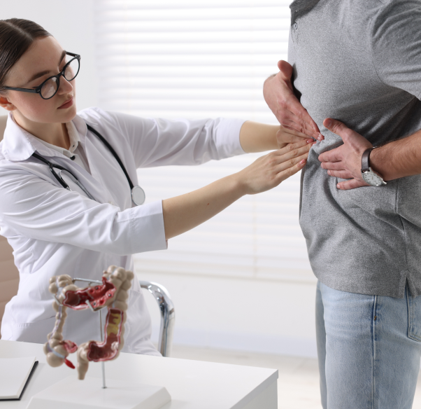
The food you eat is carried from the throat to the Stomach by a tube called the Oesophagus. Achalasia is a rare condition in which your Oesophagus is affected. Achalasia is also called Achalasia cardia. It affects 1 in every 100, 000 individuals. The Lower Oesophageal Sphincter (LES) is an opening (valve) that opens into your Stomach. The food that you eat is pushed into the Stomach by the opening of LES. But, if you have Achalasia, your LES fails to open during swallowing. This leads to a pile-up of food within your Oesophagus. Most commonly, this condition can be a result of damage to your Oesophageal nerves, or due to the damage to LES.
Symptoms
The inflammation causes swelling and irritates the intestine to cause it to contract faster hence causing Diarrhoea.
- Bleeding in stool
- Weight loss
- Bone pain
- Skin problems
- Anal Fissures
- Fever
- Persistent and excessive bleeding can lead to Anaemia
- Abdominal pain
- Mouth sores
In children, malabsorption due to digestive problems can lead to arrest in growth and development. The disease has a waxing-waning character and relapses occur in several episodes throughout life.
Complications may develop in the course of the illness. The inflammatory patches may heal to form scars that contract to cause strictures which can narrow the lumen of the affected segment and carry a risk of obstruction.
The Ulcers may deepen to form blind channels called fistulae that open into the lumen or externally on the skin of the anus. The lining of the lumen may develop deep and large cracks called fissures that are very painful.
Diagnosing Crohn’s Disease
A thorough physical examination and a series of tests are required to diagnose CD.
You may be advised a blood test which may reveal Anaemia, and an increase in the counts of white blood cells which signifies inflammation. Blood tests may be done to measure the tiers of specific proteins produced by specialised cells of the active immune system.
Some of these include Tumour necrosis factor α (TNFα), C reactive protein (CRP), and antibodies like perinuclear anti-neutrophilic cytoplasmic antibody (P-ANCA), and anti-Saccharomyces cerevisiae antibodies (ASCA). Antibodies are specific proteins produced by white blood cells as a part of the immunological response.
A series of tests for liver function may also be done.
A stool test may reveal bleeding and inflammation.
You may be required to undergo a series of X-rays after drinking barium meal, a viscid solution that will coat the digestive tract and help to localize the inflammation in the upper GI Tract. A barium enema may be administered to localised inflammation in the lower GI Tract.
Besides these, your doctor may want to do an endoscopy of your GI Tract to visually inspect the affected part and obtain a fragment of tissue for a detailed microscopic examination. A flexible lighted tube mounted with a small camera and linked to a TV monitor is inserted up the anus to inspect the intestine.
Red patches with Ulcers and bleeding can be visible in endoscopy.
Treatment for Crohn’s Disease
The Crohn’s Disease treatment includes medicines, nutritional supplements, and surgery. One or more options may be chosen depending upon the location, severity, complications, and response to previous treatments if being treated for recurring symptoms.
Medicines like mesalamine are used to control inflammation. These are not absorbed through the GI Tract and the anti-inflammatory agent is released to act locally in the GI Tract. Another class of medicines called steroids like prednisone and immune-suppressive agents like 6-mercaptopurine, and azathioprine are used to alleviate the response of the immune system and hence control inflammation. When these options fail, another medication called infliximab which is anti-TNFα is used to block the body’s immune response.
Antibiotics may be used to control the bacterial overgrowth in the intestines. Fluid replacement is required in dehydration due to Diarrhoea. Around 70-80% of patients may ultimately need surgery to control symptoms or complications like perforation, stricture, or excessive bleeding. Nutritional supplements are used in children and in those who show nutrient deficits.
The goal of treatment is to control inflammation, relieve symptoms, and correct nutritional deficiencies. CD cannot be cured, though the number of episodes can be reduced. When the disease flares, the treatment required to arrest inflammation may not be the same in all episodes.
Surgery is advised when medical treatment fails or in case of complications. Surgery cannot eliminate the disease as inflammation may recur in the previously healthy area after surgery. Multiple surgeries may be required in some patients who are suffering from symptoms of Crohn’s Disease. A part or whole of the large or small intestine may be removed during surgery and one may need to do good care of the surgical stump.
Essential nutrient deficiencies may vary from person to person depending upon the segment and extent of involvement of the GI Tract. Adequate nutritional supplements can help to prevent essential nutrient deficiency.
Inflammatory Bowel Disease is characterised by inflammation in the digestive system, also called the Gastrointestinal (GI) system. The inflammation is mediated by the Immune System which is the defence system in our body and fights invading disease-causing germs. This system may become erroneously active and attack the indigenous resident flora of the gut or a layer or structural molecule in the GI system. The resultant inflammation is characterised by redness, swelling, ulceration, and tissue destruction in the affected part(s).
It affects around 4 to 10 people per 100,000 population per year. This entity may need specialized management and patient compliance.
Crohn’s Disease (CD) is a type of Inflammatory Bowel Disease. The inflammation extends deep into the wall of the affected part and can be found in any part of the GI system from the mouth to the anus. Most commonly, the last part of the Small Intestine called the Ileum, is affected.
The inflammation is patchy, i.e., segments of the normal bowel wall are found between the multiple loci of inflammation.
Signs and symptoms
Signs and symptoms of Anal Fissure include:
- Sharp, burning pain while passing stools, that often lasts for a longer time
- Bleeding when you pass stools
- Itching
- Reluctant to have a bowel movement (may lead to faecal impaction)
- Discharge of pus from the fissure
Causes and risk factors
Anal Fissures are caused by the trauma to the inner lining of the anus. The condition occurs due to passing dry or hard stools, straining during childbirth, tight anal sphincter muscles, or frequent bowel movements.
Anal Fissures may also develop due to conditions such as Anal Cancer, Herpes, HIV, or Syphilis.
Anal Fissure Treatment
Mostly, Anal Fissures heal without the treatment. But if the condition becomes chronic, you may require interventions to promote healing.
Complications
Complications include recurrence and extension of the tear to the surrounding muscles. If the Anal Fissure fails to heal within six weeks, then the condition is chronic.
When to seek medical advice?
Anyone of us can experience Anal Fissures, so don’t let embarrassment stop you from seeking medical advice. Consult your doctor if you have severe pain during a bowel movement, or you notice blood on stools after a bowel movement or if the fissure persists for more than six weeks.
Diagnosis
Diagnosis of Anal Fissures includes a visual examination of the anal area and a digital rectal exam. A digital rectal exam is performed by inserting an endoscope into the rectum to visualize the tear easily.
Generally, Anal Fissure appears as a paper cut. In the case of a Chronic Fissure, a tear will be present along with two lumps or tags of skin, one internal and one external. The internal lump is called the sentinel pile, and the external lump is called the hypertrophied papilla.
Non-surgical treatment
The goal of non-surgical treatment is to reduce the pain and make the stool soft for easy bowel movement.
- Using stool-softeners
- Applying nitro-glycerine ointment
- Applying topical anaesthetic creams such as lidocaine
- Taking sitz baths to relax anal muscles
- Treating with Botox (Botulinum toxin type A) injection to prevent spasms in the anal muscles by paralyzing them
Surgery
Surgery is indicated in chronic Anal Fissure treatment and for cases that cannot be cured with other treatments, then your doctor opts for surgery. Lateral Internal Sphincterotomy (LIS) is the common surgery performed to treat Anal Fissures. The procedure involves cutting a small part of the anal muscle to reduce spasms and promote healing.
Inflammatory Bowel Disease or Anal Tumours also have similar symptoms. So, your doctor may further evaluate the condition if the fissure doesn’t heal with the treatment.
Prevention
Fissures in the anus can always be prevented by following some simple tips:
- Cleanse the anal area with warm water and mild soap
- Drink plenty of fluids
- Consume fibre-rich diet
- Perform physical activity regularly
- Treat conditions such as Constipation and Diarrhoea immediately
In the case of infants, change the diapers frequently and keep the anal area dry.
One in every ten people can be affected with Anal Fissures in their lifetime. These are cracks or tears in the lining of the lower rectum (anus). They do not cause serious problems and are not life-threatening. The condition can occur at any age, but most seem in infants and young children.
Symptoms of Aphthous Ulcer
- Formation of a small, oval-shaped, white, or yellow coloured Ulcer
- Painful red area surrounding the Ulcer
- Tingling sensation in the mouth
Swollen lymph nodes, fever, and weakness are found in some cases. Also, the Canker Sores or Aphthous Ulcer heals without scarring in one or two weeks. Then your doctor may suggest certain laboratory tests to rule out other health conditions such as Cancer.
When to seek medical advice?
You must visit the doctor if you have:
- A large sore
- Recurring sores
- Persistent sores that do not heal within one to two weeks
- Extreme pain that makes eating and drinking difficult
- High fever
Diagnosis of Aphthous Ulcer
Your doctor may identify the Aphthous Sores with a visual exam. Therefore, no other test or diagnosis is required for Canker Sores. But if you have persistent or recurrent Canker Sores.
Causes and risk factors
The cause of Aphthous Ulcers remains unclear. The combination of certain triggering factors may lead to the formation of these Ulcers.
- Injury: An injury to your mouth from dentures or an accidental cheek bite
- Hormonal changes: In women, Aphthous Ulcers may occur during menstruation due to hormonal changes.
- Genetics: A genetic predisposition may also cause Aphthous Ulcers.
- Nutritional deficiency: Lack of iron and vitamins such as vitamin B12 and folic acid may cause Ulcers in the mouth.
Other causes include food allergies, medications (nicorandil, anti-inflammatory medicines), smoking, viral and bacterial infections, and emotional stress.
Treatment for Aphthous Ulcer
There is no treatment for Aphthous Ulcers. But there are certain medications which tend to relieve the symptoms. For minor Aphthous Sores, treatment may not be required. If the sores are larger and persistent, your doctor may prescribe the following for Aphthous Ulcer treatment:
- Mouth rinse - containing dexamethasone helps to reduce pain and inflammation.
- Topical applications - such as benzocaine, fluocinonide, and hydrogen peroxide may help relieve pain and promote the healing process.
- Oral medications - such as steroids and vitamin supplements may be given.
Self-Management
As there is no best treatment for Aphthous Ulcer, self-management is necessary. Consider the following tips to relieve pain, promote healing, and prevent recurrent aphthous stomatitis.
- Cleanse your mouth using salt water or baking soda.
- Take a small amount of milk of magnesia and spread it gently on the Ulcer.
- Apply ice on the Aphthous Ulcers.
- Avoid spicy foods, acidic fruit juices, and salty foods.
- Practice good oral health and use a very soft toothbrush.
- Correct the nutritional deficiencies with vitamin and mineral supplements.
Aphthous Ulcers, also known as Canker Sores, are the most common type of mouth Ulcers. A report said that one in five individuals might develop Aphthous Ulcers at any stage of their life. These Ulcers in the mouth are painful and recur from time to time.
Aphthous Ulcers are painful sores that affect the lining tissues of the oral cavity. These painful sores occur anywhere in the mouth except the hard palate (roof of the mouth) or on the gums present right beside the teeth. The Ulcers appear as small, shallow lesions. These lesions cause difficulty to eat. This type of ulcer is most seen in the age group of 10 to 20 years.
Causes
The exact cause of Autoimmune Pancreatitis is not known, but it is due to the immune cells attacking your pancreatic cells, like other Autoimmune Diseases.
Risk factors
Type I AIP
Gender: The condition is predominant in males when compared to females
Age: Occurs in your 60’s and 70’s
Type II AIP
Gender: The condition is equally likely in both males and females
Age: Occurs in your 40’s and above
Complications
If a timely treatment is not made available for the patient with Autoimmune Pancreatitis, then the following complications can occur:
Diabetes: In Autoimmune Pancreatitis, insulin production is affected due to the damage to the Pancreas. This can lead to Diabetes, and the patient may require oral Anti-diabetic medications or insulin injections.
Pancreatic insufficiency: As the ability of the Pancreas to produce adequate enzymes is affected in AIP, the patient may have Diarrhoea, weight loss, vitamin or mineral deficiency, and metabolic bone disease.
Pancreatic calcifications or stone formation can occur in the Pancreas over time.
Treatments such as long-term steroids used for AIP management also can cause certain complications. However, the life expectancy of the patients treated is normal.
Diagnosis
An accurate diagnosis of Autoimmune Pancreatitis is important as it has similarities with Pancreatic Cancer. Underdiagnosis can lead to a delay in treatment or a wrong treatment.
Type I AIP
You have type I AIP if your diagnostic tests show:
- One or more masses in the Pancreas or occasionally in the other Organs
- Elevated levels of IgG4 (antibodies) in the Pancreas or occasionally in the other Organs
AIP type II
You have type II AIP, if your diagnostic tests show:
- Normal levels of IgG4 (antibodies) in the Pancreas or the other organs
- Granulocyte epithelial lesions in the pancreatic ducts which destroy the ducts
An Endoscopic biopsy must be done for a definitive diagnosis of type II AIP. During the biopsy, the distinctive appearance of the pancreatic tissue is identified under the microscope which indicates type II AIP.
Treatment
Before the Autoimmune Pancreatitis treatment begins, a biliary stent is inserted to drain the bile from the ducts in case obstructive Jaundice is identified.
Steroidal therapy: Autoimmune Pancreatitis is highly responsive to steroidal therapy. It regresses or resolves with treatment using steroids. A short course of prednisolone improves the symptoms of AIP. People with type I respond faster to the treatment with steroids compared to those with type II. However, the chances of relapse after the treatment are more in type I.
Steroids also can treat extra pancreatic manifestations which need continuous monitoring.
Immunosuppressants or immune modulators may be used in case of relapse of AIP which is possible in 30-50% of the patients treated with steroids. As long-term treatment with steroids can lead to serious side effects, immune-suppressing agents such as mercaptopurine, azathioprine, mycophenolate, and rituximab are given.
For patients with pancreatic insufficiency, supplementary enzymes are given.
If Diabetes has occurred as a complication of AIP, then appropriate anti-diabetic medication will be given.
Autoimmune Pancreatitis Symptoms
The signs and symptoms of Autoimmune Pancreatitis and Pancreatic Cancer are similar, but the treatments are different for both. Therefore, it is important to distinguish them.
Autoimmune Pancreatitis symptoms are usually not evident but include the following in the patients who have symptoms:
- Yellow skin and eyes (painless Jaundice which is the most common presenting symptom)
- Amber-coloured urine
- Nausea, vomiting
- Weakness and fatigue
- Chronic and recurrent pain in the abdomen
- Pain in the middle part of your back
- Pale stools or stools that float
- Bloating and loss of appetite
- Unexplained weight loss
- Enlarged Pancreas, which is difficult to differentiate from Pancreatic Cancer
- Strictures in the pancreatic duct
- Extra pancreatic manifestations such as enlargement of salivary glands and lymph nodes, scars in the bile duct, kidney diseases, and liver inflammation
Autoimmune Pancreatitis (AIP) is a rare, but chronic inflammatory disorder of the Pancreas. It is caused due to the body’s own defined mechanism attacking the Pancreas. They are of two types namely, type I AIP and type II AIP. Type I affects multiple organs along with Pancreas while type II affects only the Pancreas.
Understanding Celiac Disease
Celiac Disease is an Autoimmune Disease. The defined system of the body called the immune system which reacts to invading disease-causing germs mistakenly recognizes the intestinal enzyme called tissue transglutaminase (tags) as harmful and mounts an attack causing inflammation.
Our intestines have villi, or thin finger-like protrusions in the luminal surface which help in absorption of the digested food. Due to the immune attack, these villi get flattened and do not aid in absorption. In the long term, it causes malnourishment as it hampers the absorption of major nutrients from the intestines into the blood.
Gluten, a protein found in wheat, rye, triticale, and barley, is the culprit. It triggers the immune response in patients with Celiac Disease. Such people are also intolerant to other gluten-containing products like lip balms and some medicines.
Celiac Disease is a genetic disease meaning that it runs in families. You may first get to know of it after surgery, pregnancy, childbirth, viral infection, or severe emotional stress. You may have or may be at risk of developing other autoimmune disorders like Diabetes type 1 or thyroid disorders.
You may need nutritional supplements to make up for the deficiencies the disease has caused in you. No other medication will usually be required. Rarely, you may need steroids to suppress the immune activity.
A drug called dapsone may help in dermatitis herpetiformis.
Living with Celiac Disease
Celiac Disease is a serious condition. You may develop deficiencies in various essential nutrients like vitamins and minerals. You may develop Anaemia due to deficiency of iron and show easy bruising and bleeding due to deficiency of vitamin K. It can affect numerous organs like the liver, Pancreas, gallbladder, bones, heart, nervous system, etc.
You may have fertility problems. Lastly, you may stand a risk of developing Cancers in the digestive system.
Celiac Disease cannot be ignored. There is no cure for Celiac Disease. The best bet is to remain on a gluten-free diet. As the intestine heals, symptoms both trivial and severe, respond dramatically to gluten restriction. Change your food habits and learn to avoid even small amounts of gluten. Read the food labels carefully.
Beware! Wheat-free is not gluten-free. It may have other grains that you may need to avoid. These include rye, barley, einkorn, kaput, spelt, and triticale.
You may need to avoid bread, broth, commercial cereals, wafers with grains, pasta, cakes, marinades, sauces and stuffing, vegetable gums, food starch, dextrin, etc. Avoid cross-contamination with gluten products. Keep a separate toaster for yourself. Do not use a knife that has been used to spread jam or butter on a bread slice. Buy your food in a bakery that has separate gluten-free machinery.
You can enjoy fruits, vegetables, eggs, corn, rice, buckwheat, sorghum, arrowroot, garbanzo beans, chickpeas, quinoa, tapioca, teff, and potatoes, meat, fish, chicken, legumes, nuts, seeds, oils, milk, and cheese!
Symptoms
Celiac Disease may manifest a variety of symptoms either related to the digestive system or other systems. Symptoms may be deceptively absent until late adulthood. The commonest Celiac Disease symptoms are:
- Diarrhoea
- Bloating
- Abdominal pain
- Stools may be frothy due to unabsorbed fats
- Anaemia
- Unexplained weight loss
- Skin rash
- Depression
- Brittle bones
- Bone and joint pain
- Mouth Ulcers
- Fatigue
- Tingling or numbness in hands and feet
- Infertility
15-25% of patients with Celiac Disease may have an itchy and blistering skin rash on areas like elbows, knees, and buttocks. The rash may be the sole sign of Celiac Disease and may not be accompanied by digestive or other complaints. This condition is called dermatitis herpetiformis (DH).
Babies and children may have the following symptoms:
- Pale and deficient in red blood cells
- Anaemia
- Diarrhoea
- Bloating
- Abdominal pain
- Weight loss
- Delayed puberty
- Stunted growth
- Irritable
- Behavioural problems
Diagnosing Celiac Disease
Celiac Disease is diagnosed by detecting antibodies in blood. These are proteins produced by the hyperactive immune system.
You may need to undergo an endoscopy wherein a flexible tubing with a camera-mounted tip would be inserted into the digestive system to view the interior and retrieve a piece of your intestine for a detailed microscopic and laboratory examination.
Gene analysis is rarely done. You should also have your family screened for Celiac Disease.
Your doctor may require you to take a gluten unrestricted diet for a few days before you undergo tests to facilitate the Celiac Disease diagnosis.
Whenever you go for a family dinner, you must order separately for your wife. She is intolerant to wheat-based products. Whenever she consumes anything made of wheat, she develops abdominal pain and Diarrhoea. It becomes quite difficult to choose foods that do not have a wheat base. Most of the time, she orders rice-based foods, vegetables, and fruits. Eating out becomes quite a challenge!
The condition is called Celiac Disease, also called celiac sprue, no tropical sprue, and gluten-sensitive enteropathy. A change in lifestyle and precaution can help you to keep up the quality of your life and have good health.
Symptoms
Below are some common symptoms of the infection.
- An infection with C. difficile causes illness of varying severity according to the strain of bacteria predominantly inhabiting the gut.
- Increase in bowel frequency leading to a watery Diarrhoea,
- Fever
- Feeling of vomiting
- Abdominal pain
- Lack of appetite
- Weight loss
A more severe form of illness is pseudomembranous colitis, where the colon is inflamed, and the inner lining develops membrane-like patches. This causes bloody Diarrhoea, crampy pain in the abdomen, fever, and swelling and distention of the abdomen. The colon may distend, and the wall progressively thins down to cause a rupture. This is called a perforation.
This can be life-threatening as the contents of the colon can spill over into the abdomen and cause a widespread infection. It may sometimes lead to death.
A transient period of Diarrhoea is common after antibiotic use in many people.
One should see a doctor when the stools last more than 3 days, become more frequent, or are accompanied by blood or pus. Fever, cramping, feeling of fullness or vomiting, or weight loss due to a non-remitting Diarrhoea are other reasons to seek medical help.
Diagnosis
C. difficile infection is suspected in anybody who develops Diarrhoea and has taken antibiotics for any reason in the past two months or when Diarrhoea develops after hospitalization.
The doctor may then like to do some tests to confirm the diagnosis. The bacteria can be detected in a stool sample by using several laboratory tests like enzyme immunoassay, polymerase chain reaction and tissue culture assay. So, stool testing is a simple way to detect the infection.
In the more severe cases, a computerized tomography (CT) scan may enable examination of the colon through a series of images. The colon wall may be thickened which may be suggestive of pseudomembranous colitis.
The inside of the colon may be directly viewed through a flexible sigmoidoscopy. A tube with a camera-mounted tip may be inserted through the anal opening upward to look at the interior of the colon. This may reveal areas of inflammation and patches of membranes.
Treatment
The first essential requirement in treatment is to discontinue the intake of a suspect causative antibiotic. In very mild cases, this alone may be curative. Many people may need further treatment.
Paradoxically, the treatment then involves the administration of another antibiotic that acts against C. difficile and controls infection.
The antibiotics used for this purpose are metronidazole for moderate cases and vancomycin for more severe cases. The two are taken by mouth and arrest the growth and proliferation of C. difficile and help restore the normal bacterial colonies in the gut.
In an extreme event of inflammation and organ failure, surgery may be done to remove the diseased segment of the colon.
Probiotics are another option. These are bacteria or yeasts that help to restore the natural and normal flora in the gut. These can be taken along with antibiotics in the treatment of C. difficile. Probiotics also help to prevent recurrent disease.
Episodes of infection may recur as the previous ones may not have recovered fully or a reinfection may occur with a different strain. In this case, the antibiotics and probiotics are given for longer durations. In any case, it is important to provide adequate fluid replacement. Some doctors may not like to add probiotics.
One should take frequent small sips of fluids to prevent dehydration. In more severe cases, fluids may need to be infused through the blood vessel.
Medicines that decrease the motility of the gut to control Diarrhoea, such as loperamide, should be avoided as these delay the clearance the toxins from the gut and may make the illness more grievous.
Prevention
Infection with C. difficile can be prevented.
- The foremost precaution is to judiciously use antibiotics. This class of medicines should only be taken for confirmed bacterial infections and their use in viral illnesses should be avoided. In addition, these should be taken for the prescribed number of days and in the doses as advised by the healthcare personnel.
- Extended and irrational use of antibiotics is always harmful.
- The hospitals, nursing homes, and other healthcare facilities should be kept clean.
- The caregivers should be trained to implement precautions.
- Hand washing can prevent many episodes of C. difficile infection. Each healthcare worker should thoroughly wash hands with soap and water after examining every patient and before handling the next patient.
- The patients should be instructed to wash their hands before and after the use of the bathroom.
- Hospital staff and visitors should wear protective masks and gloves.
- All surfaces, floors, bedpans, furnishings, and equipment should be cleaned and sterilized.
- Chlorine bleach is an effective disinfectant for the elimination of C. difficile, both the active and the dormant forms of the bacteria.
Clostridium difficile (C. difficile) is a bacterium that is normally present in the gut of healthy individuals and does not cause any problems. When the flora in the gut is disturbed due to inappropriate use of anti-infective medicines called antibiotics, the bacteria multiply to increase in numbers and predominantly populate the gut. The person is then said to be ‘infected’ with C. difficile. The bacteria then produce ‘toxins’ that cause Diarrhoea, and other problems like inflammation of the Large Intestine, called colitis.
Anybody can develop an infection with C. difficile though the elderly are more prone than the youngsters. The commonest risk factor is the intake of antibiotics for the treatment of any medical condition. The risk is greater if a broad-spectrum antibiotic is used for a prolonged period or multiple antibiotics are used together.
Most infections occur in hospitals and nursing homes where the bacteria spread easily, and people are more vulnerable. Person-to-person spread mainly through hands is known to occur in such settings.
Toilets, stethoscopes, thermometers, bedrails, bedside tables, and other furniture or equipment may facilitate the spread if contaminated with the bacteria. Thus, anybody who has been hospitalized in the recent past is at risk.
People with compromised defences of the body and with a weak immune system are prone. This includes patients on treatment for Cancers. One who has had an infection with C. difficile in the past is at risk for more infections in future. So are the patients with Cancer or inflammatory disease of the colon.
Identifying Constipation
When you are constipated, besides not able to defecate completely you will feel full and have a reduced appetite.
During bowel movements, you may have a hard time emptying your Stomach, straining too much will hurt a little on the go.
You may find drops of blood on the skin of the anus.
Your belly may look a little bulged out due to hard and solid faecal matter, which doesn’t defecate easily.
After you are done you may still have the urge to go again. If you think you are constipated, visit your doctor immediately.
Treating Constipation
Consuming a fibre-rich diet is best during Constipation. It is said that men should consume about 38 grams of fibre every day and women should consume about 25 grams of fibre per day.
Make it a point that you include fruits and vegetables in your daily diet. Eat cereals that contain bran or use brands as toppings on your food. If you are starting to include fibre in your diet, then start slowly and increase gradually to avoid bloating and gas formation. Also, remember to drink plenty of water.
Prevention
A few simple steps to prevent Constipation are
- Eat more fibrous food
- Drink plenty of fluids, which may include water, juice, soups, or any other drink
- Exercise regularly
- Say NO to alcohol and caffeine as they deplete the water content in the body
- Never ignore an urge to have a bowel movement
- Allot plenty of time for a good and relaxed bowel movement
- Do not take too much of laxatives as it can worsen your condition
- Avoid foods that are high in fat and sugar as they may cause Constipation
Constipation in children
Constipation is a very common problem among children. A constipated child has hard and dry stools and it is painful for the child to pass bowel movements.
Treating Constipation
Although treatment procedure differs depending on the severity and duration of Constipation, dietary changes help to relieve the symptoms.
Treatment includes eating food like whole grains, bran cereals, fruits, vegetables, and other fibre-rich food, and consuming enough water, fruit, and vegetable juice to avoid dehydration.
In addition, you should allot time for regular exercise.
Do not ignore the urge to have a bowel movement. In severe cases, laxatives and enemas may be recommended for a short time
Diagnosis
Diagnosis is done based on the severity, age, duration, whether blood is present in the stool, changes in bowel habits, or if weight loss has occurred. Most extensive tests are not required to diagnose Constipation, medical history and a simple physical examination are all that is needed to identify Constipation.
A detailed medical history may include information like duration of Constipation, consistency of stool, frequency of bowel movement, and whether blood strains are found in stool. Your doctor may also ask about your eating habits, medications, and the amount of physical activity, all these will help him to determine the exact cause of Constipation.
The physical examination includes a rectal examination with a lubricated and gloved finger to check the muscles around the rectum for tenderness, obstruction, and blood strains.
Other extensive tests are usually done in people with severe symptoms. These include performing tests like – Sigmoidoscopy to check the Large Intestine; colonoscopy to investigate the entire Large Intestine; colorectal transit study where you swallow a small capsule and the path of the capsule through your intestine and anus is seen using an X-ray study; a no-rectal function test & defecography to see how effectively you can push your faeces out.
Using laxatives and enemas
Using laxatives is not recommended so try to minimise its use. They should not be used for a long term of period. But bulk-forming laxatives are the exception to this. Bulk laxatives function naturally to add water to the stool so that they are defected easily.
Bulk-forming laxatives can be used daily either separately or you can add them to your juice and drink. Start with a mild dose and gradually increase the dosage every 3 to 5 days until defecation becomes easy.
Remember to drink plenty of water.
Bulk-forming laxatives may sometimes cause side effects like bloating or gas formation, but this goes away in a few weeks. Many wonder whether they can use mineral oil as a laxative, you can but only when your doctor prescribes it.
He might recommend the use of mineral oil in case you had surgery and should not strain yourself during bowel movements. But the major side-effect of using mineral oil is that it causes a deficiency of vitamins A, D, E, and K. Using an enema doesn’t prove effective for Constipation and long-term use of both laxatives and enemas is not recommended.
What causes Constipation?
The process of digestion starts once you start munching your food. It passes through your mouth, Stomach, and Small Intestine, then heads towards the Large Intestine, and finally out of the body through the rectum.
Constipation occurs when your Large Intestine absorbs too much water from the food making it hard and difficulty to push it out of the body. The various causes of Constipation are.
- Lack of fibre in the diet
- Some medications for Constipation are painkillers and anti-depressants
- Taking calcium & iron supplements
- Irritable bowel syndrome
- Milk
- Change in routine activity due to pregnancy, travel, or ageing
- Abuse of laxatives
- Not drinking enough water/dehydration
- Not responding to the urge to have a bowel movement
- No physical activity
- Problem in colon and rectum
- Problem with intestinal function
- Stroke
- Stress
Foods rich in fibre
Foods that are rich in fibre are listed below
| Cereals | Whole-grain cereal, black-eyed peas, kidney beans, wheat or grains bread, oats, maize, barley, whole-wheat |
| Fruits | Apple, peach, Avocado, guava, apricot, raspberry, fig, date, gooseberry, mango, orange, pear, wood apple, jamun, papaya, pomegranate, plum, peach, kiwi fruit and grapes. |
| Vegetables | Squash, turnip, lady’s finger, radish, broccoli, beans, brussels sprouts, cabbage, carrots, cucumber, broccoli, peas, onion, tomato, brinjal, cauliflower, bitter guard, spinach, beetroot, sprouts |
Include these foods in your daily diet for a healthy Stomach.
People of all ages suffer from Constipation. Constipation means that you are not passing your stool (faeces) as often, and as normally you do. During Constipation, you might have to strain more than usual or be unable to empty your bowel. Your stool becomes hard, dry, and small which is difficult to eliminate. Constipation usually lasts for a short period with no long-lasting health effects.
Many people think that they are constipated if they do not have bowel movements every day. However, the number of bowel movements per day depends on the person, some may have a normal bowel movement of 3 times a day and for some, it may be 3 times a week. Normal bowel movement depends on the food that you consume; how active you are and many other factors.
Types
Common types of Diverticular Disease are
- Diverticulosis
- Diverticulitis
- Diverticular bleeding
Diverticulosis: Diverticulosis is the term for the presence of more than one diverticulum in the large bowel. Infection may develop when bacteria or stool gets caught in the Diverticula. This can result in rupture of the colon, and infection or collection of pus in the tissues that surround the colon. This condition is termed as Diverticulitis.
Diverticular bleeding: Diverticular bleeding is when the wall of the colon bleeds due to the development of a Diverticula on the colon.
Symptoms
The majority of the patients with Diverticulosis may have few or no symptoms. It may be a chance finding during an Endoscopic X-ray examination. A few of the Diverticulitis symptoms are:
- Pain and discomfort in the left lower abdomen
- Bloating, or change in bowel habits (Constipation/ Diarrhoea)
- Severe and constant pain over the left lower abdomen
- High temperature
- Vomiting
- Abdominal tenderness
- Constipation/Diarrhoea
There is acute bleeding from the rectum in cases with Diverticular bleeding. This may be associated with shock-like symptoms and dizziness depending on the amount of blood loss.
The complications of Diverticulosis are Diverticulitis, bleeding into the colon, and colon obstruction. A diverticulum can also rupture and bacteria within the colon can spread to the tissues surrounding the colon. The collection of pus around the inflamed diverticulum can lead to the formation of what is called an abscess.
Risk factors
Your risk of developing Diverticular Disease increases with
- Advancing age
- Obesity
- Family history
- Chronic Constipation
- Lack of exercise
- Frequent use of laxatives
It is very unlikely that you will get the disease before the age of 40. It is most common after the age of 60. Both men and women are equally affected.
Diagnosis
Your doctor may diagnose Diverticular Disease by performing one or more of the following tests. A barium enema (X-ray test performed with injection of liquid material into the colon through the rectum) will help in visualizing the anatomy of the colon, and identify if Diverticula, polyps or growths are present. Your doctor may visualize the inside of your colon by colonoscopy with the aid of a thin, flexible tube with a light and camera and look for Diverticula as well as polyps and other growths.
A CT scan (X-ray test that takes multiple section pictures of the body) can help to identify Diverticula but is generally not performed to make a diagnosis of Diverticular Disease.
Prevention
It is not known if you can prevent Diverticular Disease. Having a diet rich in fibre (minimum of 15 to 30 grams in a day), restriction foods with indigestible particles such as popcorn, nuts, and fruits with small seeds, drinking plenty of fluids, and exercising regularly are a few measures in the prevention of Diverticular Disease.
While Diverticulosis may be asymptomatic in many patients, the development of complications such as infection, pus formation, bleeding or obstruction of the colon may mandate hospitalisation and effective medical and surgical management. Once a diverticulum develops it does not go away. So why not try to prevent it in the first place? Increasing fibre intake in the diet, preventing Constipation, exercising regularly, drinking plenty of fluids, and avoiding excessive laxatives can help protect your large bowel from the disease condition.
Treatment
Diverticulitis treatment depends on how serious the problem is and whether you are suffering from Diverticulosis or Diverticulitis. But it is better to be aware that once a diverticulum has formed in your colon, it will not go away. If you do not have symptoms of Diverticulitis, then you may try to increase the fibre in your diet to soften and bulk the stool, drink more fluids, and exercise regularly to prevent the development of more Diverticulitis or associated complications.
High-fibre foods include beans and legumes, bran, whole wheat bread, whole grain cereals, fruits (apples, bananas, and pears), vegetables such as broccoli, carrots, corn and squash, brown rice and whole wheat pasta. Laxatives in consultation with your doctor may help in avoiding Constipation.
Treatment of complications
Mild cases of Diverticulitis may be managed with oral antibiotics, dietary restrictions, and stool softeners. More severe cases require hospitalisation with intravenous antibiotics and dietary restraints. If there is pus collection around your colon, then your doctor may need to drain it by a catheter passed into your abdomen through the skin and guided by radiologic support.
Acute bleeding due to Diverticulosis requires in-hospital management with intravenous fluids, blood transfusions, and diagnostic procedures to identify the site of the bleeding. If you do not respond to medical management and have recurrent episodes of Diverticulitis, obstruction of the colon, or severe bleeding from the Diverticulum, Diverticulitis surgery may be necessary to remove the involved area of the colon. Or else a temporary opening for your colon may need to be done on the abdominal wall.
Diverticular Disease affects the colon which is the part of your large Intestine. It stores and eliminates waste material from your body. In this condition, there is a formation of small pouches or sacs in the lining of your bowel, particularly the large Intestine. The bulging sac or pouch is called a diverticulum. More than one diverticulum is called a Diverticula. These sacs are seen to occur more commonly near the lower end of the left colon (sigmoid colon). The condition of having these Diverticula in the colon is termed sigmoid Diverticulosis.
While it is not certain why Diverticulosis develops, there are a few theories that have been suggested. With advancing age, the muscular wall of your colon becomes thicker, although there is no clear understanding of the cause for this thickening. The resultant abnormal contraction and spasm can cause intermittent high pressure in the colon. This increased pressure within the colon causes bulging pockets of tissue or sacs that push out from weak spots in the colon wall.
Another theory suggests the role of low-fibre diets or diets that are low in roughage in the development of Diverticula. Without fibre, the stools are small and dry and are difficult to pass. Intestinal muscles need to contract with greater pressure to force the stools along. This allows for segments of the colon to close off from the rest of the colon when the colonic muscle in the segment contracts. The pressure within the closed-off segments becomes high as the increased pressure cannot dissipate to the rest of the colon. Over some time, high pressures in the colon can push the inner intestinal lining outward through weak areas in the muscular walls forming pouches. The other factors implicated are chronic Constipation and lack of exercise.
Types
Dysphagia is broadly classified into two types; oropharyngeal Dysphagia and Oesophageal Dysphagia. In oropharyngeal Dysphagia, you will have trouble moving food from your mouth into your upper Oesophagus . Trouble moving food through your Oesophagus into your Stomach is termed as Oesophageal Dysphagia.
Causes
Poor eating habits such as eating too fast, taking large bites, eating while lying down, and not drinking enough water while eating can cause Dysphagia. It can happen if you have difficulty chewing your food due to missing teeth or dentures. You may also have Dysphagia if you suffer from acid reflux which can cause scar tissue and narrow the opening of the Oesophagus.
Diseases of the muscles or nerves that control the muscles of the pharynx, and Oesophagus or damage to the swallowing centre in the brain (area of the brain that controls the act of swallowing) can cause Dysphagia. Disorders such as a paralytic attack, degenerative disorders of the brain such as Parkinson’s disease or multiple sclerosis, and muscle-wasting diseases such as myasthenia gravis can stop the nerves and muscles in your Oesophagus from working right. This can result in food moving slowly or even getting stuck in the Oesophagus.
Diseases specific to the Oesophagus such as Achalasia (valve at the lower end of the Oesophagus fails to open and let food pass into the Stomach), eosinophilic Esophagitis (inflammation of the Oesophageal wall), and tumour growths in the Oesophagus can cause Dysphagia. Also, external pressure due to Cancers in the chest cavity, an enlarged thyroid, or an enlarged heart may put pressure on the Oesophagus and cause Dysphagia.
Treatment
Your doctor will decide on the best Dysphagia treatment option for you based on the cause, the seriousness, and the complications, if any. You may be advised on the need to improve your ability to swallow by following these steps if your Dysphagia is due to poor eating habits.
- Chew carefully
- Drink more water while eating
- Change the positions while swallowing
You may be advised on the intake of foods that are easy to chew. You may be taught exercises that can help strengthen your swallowing muscles. You may need medicines such as antacids or acid reducers if your Dysphagia is due to acid reflux. A medication called botulinum toxin is used if Dysphagia is caused due to problems with the muscles.
Surgery may be necessary if the Dysphagia is due to a tumour or something blocking the Oesophagus. If there is complete obstruction emergent upper endoscopy is essential. If a stricture, ring, or web is found in the Oesophagus careful endoscopic dilation is performed.
For Oesophageal Dysphagia involving an Oesophageal muscle that doesn't relax, your doctor may dilate you with a balloon attached to an endoscope.
Diagnosis
Your doctor will take a detailed history regarding your symptoms, their duration, and acuity of onset. He may ask you what foods or liquids you have trouble swallowing, whether there is pain or heartburn during swallowing, or if you have vomited blood anytime. He may order tests such as barium swallow and endoscopy to help confirm the diagnosis.
During the barium swallow, you will have to drink a liquid that will be monitored on an X-ray machine as it travels down your Oesophagus. It is useful in pointing out the area of blockage if any in the Oesophagus or any other problem responsible for your Dysphagia.
Your doctor may perform an endoscopy (uses a flexible tube with a light and a camera at the end of it to look inside the Oesophagus and the upper part of the small Intestine) to rule out the presence of abnormal growths, scar tissue, or other possible mechanical causes of Dysphagia. During endoscopy, your doctor may remove a tiny tissue sample (biopsy) from your Oesophagus to analyze and distinguish if you have reflux disease, infection or inflammation of the Oesophagus, or abnormal growths in the Oesophagus that is Cancerous or Non-cancerous.
Imaging scans such as computer tomography (CT scan), magnetic resonance imaging (MRI), and positron emission tomography PET scan may be part of the investigations for Dysphagia.
Symptoms
If you suffer from oropharyngeal Dysphagia, then these are a few Dysphagia symptoms you may experience. There may be difficulties during:
- Swallowing
- Choking sensation
- Cough while swallowing /li>
- Regurgitation of liquid through your nose
- Weak voice
- Weight loss
If you suffer from Oesophageal Dysphagia, then you may have the,
- Sensation of food stuck in your throat or chest
- Pressure sensation in your mid-chest area
- Pain while swallowing /li>
- Chronic heartburn
- Belching
- sore throat
Inadequate nutrition due to Dysphagia may lead to weight loss and dehydration. You are also at high risk of lung infections or pneumonia due to inhalation of food (aspiration).
Dysphagia is the medical terminology that describes the condition of difficulty or discomfort during swallowing. Problems at any of the stages of swallowing can lead to Dysphagia. The condition is seen to affect individuals at any age although the risk is seen to rise with advancing age.
Symptoms
In some cases, an Enlarged Spleen may not cause any symptoms. But, the underlying conditions that lead to splenomegaly will cause the splenomegaly symptoms.
- Pain that radiates to left shoulder (may indicate Spleen pain)
- Feeling of fullness
- Fatigue
- Easy bleeding
- Shortness of breath
- Weight loss
- Paleness
- Night sweats
Complications
If the Enlarged Spleen is left untreated, then it may lead to severe complications which include the following:
- Infections: Due to enlargement of the Spleen, the number of healthy blood cells is reduced making you more prone to infections.
- Ruptured spleen: Your Spleen is so soft that it can be damaged even with minor injuries. The risk of rupture is increased if you have an Enlarged Spleen. The ruptured Spleen causes severe bleeding and can be fatal.
When to seek medical advice?
If you have pain in your upper abdomen, then it is wise to look for medical care. When the pain is severe and worsens while you’re taking deep breaths, visit your doctor as soon as possible.
Self-management and prevention
If your Spleen is enlarged, then you must completely avoid contact sports such as football, hockey, and soccer. Wearing a seat belt would benefit you. It prevents damage to your Spleen if you’re in a car accident.
As surgical removal of your Spleen makes you vulnerable to many infections, you need to be vaccinated against the infections caused by Streptococcus pneumoniae, Haemophilus influenzae, and Neisseria meningitidis. Additionally, you must receive an influenza vaccine every year.
Treatment
Your doctor chooses a specific strategy for splenomegaly treatment based on the underlying condition that is responsible for your Spleen being enlarged. At times, if you’re diagnosed with an Enlarged Spleen causing no symptoms, then a ‘wait and watch’ approach would be beneficial. You have to visit your doctor for re-evaluation every six months or immediately after experiencing symptoms.
Splenectomy: The splenectomy is the surgical removal of your Spleen. However, the removal of Spleen may not affect your body functions.
Sometimes, Radiation Therapy would be an effective approach and best alternative to surgery. Radiation Therapy causes your Spleen to shrink and reduce the symptoms.
Diagnosis
Usually, your doctor will identify the enlargement of the liver during the physical examination that is by palpating your abdomen. To confirm the diagnosis, your doctor may order any of the following tests.
Blood tests: The blood tests involve a complete blood count that helps to determine the number of red blood cells, platelets, and white blood cells.
Computed Tomography (CT) Scan or Ultrasound: CT scan and Ultrasound help to determine the size of your Spleen.
Magnetic resonance imaging (MRI): MRI helps to check the blood flow through the Spleen.
Causes and risk factors
Several diseases and infections result in the enlargement of the Spleen. Generally, the Spleen becomes enlarged if it works excessively. So, any disease condition that damages your blood cells requires the blood to be filtered and the abnormal blood cells to be removed, this leads to the enlargement of the Spleen.
The causes of splenomegaly are the below conditions:
- Viral infections such as cytomegalovirus and mononucleosis
- Bacterial infections such as syphilis or endocarditis
- Parasitic infections such as malaria
- Cirrhosis and other liver disorders
- Haemolytic Anaemia
- Blood Cancers
- Metabolic disorders such as Gaucher's Disease
- Trauma
Spleen, the single largest mass of lymphoid tissue in your body, is located under the rib cage in the upper left quadrant of your abdomen. One of the Spleen problems is the Spleen enlargement. An Enlarged Spleen isn’t a disease itself but occurs as a result of an underlying condition. Various conditions make your Spleen enlarge. An Enlarged Spleen, also known as splenomegaly, is an increase in the size of the Spleen beyond the normal size.
Your Spleen is a part of the immune system and is considered to be a dual-purpose organ. It filters your blood by removing defective blood cells from the bloodstream. It produces white blood cells and antibodies that fight against the diseases. Your Spleen is vulnerable to a wide range of conditions as it is involved in many of your bodily functions.
Symptoms
In the early stages of Cancer, the symptoms are not evident. The Oesophageal Cancer symptoms appear only in the later stages and include:
- Difficulty and pain during swallowing
- Weight loss
- Hoarse voice
- Pain in the chest behind the breastbone
- Cough that doesn’t go away
- Indigestion
- Heartburn
Coping and support
It is a very sad thing for the patient to know that he/she has Cancer. This can also lead to Depression and loss of self-esteem. Therefore, coping is of high importance for such people. Keep trying ways to make yourself feel comfortable. Below mentioned are some helpful tips:
- Understand clearly and deeply about Oesophageal Cancer and make decisions for better care. You may learn this from your doctor, or online sources, etc.
- Keep in regular touch with your friends and family members. They can be of great emotional support and strength for you during your treatment. Talk to good listener friends and share your feelings with them.
TNM staging system
The doctors use this system to describe the Oesophageal Cancer stages. The diagnostic test reports are used to answer questions like:
- Tumour (T)-how deep is the Tumour growth, into the Oesophageal wall and surrounding tissues?
- Node (N)-has the Tumour spread to the lymph nodes?
- Metastasis (M)-has the Cancer metastasized or spread to other body parts?
Risk factors
Some types of Oesophageal Cancers are due to alcohol and tobacco consumption. The risk is much higher in people who use both of them.
Some other types of Oesophageal Cancers are related to the condition called Gastro-oesophageal Reflux Disease (GERD).
Due to the acidic rush from the Stomach to the Oesophagus, the cells of the walls get damaged and over time it can lead to the formation of malignant cells. Medical conditions such as Achalasia (a motility disease of the Oesophagus) can increase the risk for Oesophageal Cancer.
Your risk for Oesophageal Cancer also goes up as you age. Also, males are at higher risk for Oesophageal Cancer than females.
Complications
As Oesophageal Cancer advances, it can lead to certain complications as mentioned below:
- Pain: Initially, pain may not be present but is felt in the advanced stages of Oesophageal Cancer.
- Oesophageal bleeding: Bleeding with Oesophageal Cancer can be either gradual or sudden and can become severe at times.
- Oesophageal obstruction: This can lead to difficulty in passing food and drinks through your Oesophagus.
Prevention
As you have understood the risk factors for Oesophageal Cancer, you can take steps to prevent this Cancer.
- Quit smoking and drinking, or at least drink in moderation.
- Eat more healthy and colourful fruits and vegetables.
Maintain a healthy weight by aiming at losing 1 or 2 pounds each week.
Causes
Due to mutations in the DNA of the cells lining the Oesophagus, they become malignant. These cells repeatedly divide in an uncontrolled manner producing abnormal cells and thus the Tumour. The exact cause for these mutations to occur is not known.
The other causes include unusual infections with fungi, yeast, and Human Papillomavirus (HPV).
Certain foods such as beetle nuts can also increase the risk of Oesophageal Cancers.
Diagnosis
Because of no early symptoms, Oesophageal Cancer is usually diagnosed in the advanced stages.
If you present the signs and symptoms of Oesophageal Cancer, your doctor will take your medical history and family history. Following this, certain tests are performed to confirm the diagnosis. If Cancer is detected, further tests are performed to understand the stage of Oesophageal Cancer.
Test type | Purpose of the test |
Imaging tests (CT, MRI, PET) | Creates clear cross-sectional images of the Oesophageal tube using sound or radio waves, etc. To detect the exact location of the Cancer To know how far the Cancer has spread |
Barium swallow test | Barium coated Oesophagus is examined using X-rays The first test performed if swallowing difficulty is reported To detect abnormalities in the surface of the Oesophageal lining |
Endoscopy | To look at the inside of the Oesophageal tube with a camera fixed to the endoscope to diagnose Cancer and to determine the extent of the spread of Oesophageal Cancer |
Thoracoscopy and Laparoscopy | Cutting open the chest wall or abdomen under anaesthesia and observing the inside with a small camera to check if Cancer has spread and decide the benefits of surgery for that patient |
Biopsy | Examines small tissues from the Oesophagus under a microscope to ensure that Oesophageal Cancer is present |
Treatment
The Oesophageal Cancer treatment is based on cells involved in producing Cancer, the stage of Cancer, and your overall health. It helps to relieve symptoms and prevents the advancing of Cancer. The treatment options for Oesophageal Cancer are:
Surgery: Surgery is done to remove Cancer and can be used either alone or may be combined with other treatments. Surgery may be performed either to remove very small Tumours to remove a portion of the Oesophagus or to remove part of the Oesophagus and part of the Stomach.
Chemotherapy: Chemotherapy involves the use of drugs to kill Cancer cells. These drugs may be prescribed before or after surgery. In advanced stages, this therapy can help to relieve the signs and symptoms.
Radiation Therapy: This therapy employs high-energy radio waves to destroy Cancer cells. Radiation Therapy is generally combined with chemotherapy and can be given before or after the surgery.
Oesophageal Cancer is the formation of Cancer cells in the Oesophagus (the tube that connects the mouth to the Stomach). It starts within the cells in any part of the Oesophageal wall and later can spread to the other parts. Oesophageal Cancer is the sixth most common Cancer in the world; in India, it is the second and fifth most common Cancer in men and women respectively.
The Oesophageal Spasms may occur very rarely and may not require any treatment. However, a frequent or repeated Oesophageal Spasm causing blockage of food and liquids from entering the Stomach may require appropriate treatment to reduce further complications.
Types of Oesophageal Spasms
Two major types of Oesophageal Spasms include:
Diffuse or distal Oesophageal Spasms: Diffuse Oesophageal Spasms are characterized by irregular and uncoordinated squeezing of the Oesophageal muscles. This causes the food to remain in the Oesophagus and obstructs the food passage from the mouth to the Stomach. In severe cases, it may lead to Oesophageal motility disorders such as GERD, Achalasia, and Dysphagia.
Nutcracker Oesophagus: The nutcracker Oesophageal Spasms involve a coordinated squeezing of Oesophageal muscles.
Symptoms
The Oesophageal Spasm symptoms include the following:
- Chest pain that radiates to arms, back, neck, or jaw
- Inability to swallow the food or liquid
- Pain while swallowing
- Burning sensation in the chest
- Backward flow of consumed food or liquid (regurgitation)
Medical advice
Usually, no treatment is required for Oesophageal Spasm. But, if the spasms are frequent and cause severe squeezing pain in the chest, then you must immediately seek medical care.
Risk factors
The incidence of Oesophageal Spasm is very low. People aged between 60 and 80 years are at increased risk of developing Oesophageal Spasms.
It is often associated with Gastro-oesophageal Reflux Disease. The other factors that increase the risk of Oesophageal Spasms include:
- Hypertension
- Anxiety or Depression
- Intake of red wine or consumption of very hot or very cold drinks or foods
Self-management
A change in diet and lifestyle might help you to reduce the progression or prevent the Oesophageal Spasms. Consider the following measures:
- Avoid chocolate, mint, and alcohol as they worsen your condition.
- Avoid the intake of spicy foods, acidic foods such as tomatoes and oranges, and coffee.
- Avoid smoking.
- Maintain a healthy weight.
- Avoid stressful situations as stress may increase the severity of Oesophageal Spasms.
- Use of peppermint lozenge would benefit you by acting as a muscle relaxant.
Treatment
Your doctor might recommend the treatment based on the severity, frequency, and underlying cause of the condition. The Oesophageal Spasms treatment includes the following:
Medications: To manage the underlying conditions such as GERD, your doctor might prescribe proton pump inhibitors such as lansoprazole. Sometimes antidepressants such as trazodone or imipramine are suggested to reduce pain. Sildenafil, botox injections or calcium channel blockers such as diltiazem help to reduce the severity of contractions.
Surgery: The surgical procedure used to treat Oesophageal Spasm is called myotomy. The procedure involves the removal of muscle from the lower part of your Oesophagus to control the contractions. Another surgical treatment involves Peroral Endoscopic Myotomy (POEM), where an endoscope is used along with myotomy. The surgical procedure is considered only when you don’t respond to any of the treatments.
Diagnosis
Often, your doctor can identify the cause of Oesophageal Spasms by taking a medical history and performing a thorough physical examination. To confirm the diagnosis, your doctor might suggest any of the following tests.
- Endoscopy: Endoscopy is a Non-invasive Procedure used to examine your Oesophagus.
- Barium swallow: During the test, you will be asked to swallow a solution that contains barium and then X-rays are taken while you’re swallowing it.
- Oesophageal manometry: The test is used to measure the muscle contractions of your Oesophagus.
- Oesophageal pH monitoring: The test is used to determine the acid reflux.
Causes
The exact cause of Oesophageal Spasms is still unknown. However many research studies suggest that they occur due to abnormal neuronal activity that facilitates the swallowing action of the Oesophagus. Sometimes, the triggering factors such as consumption of drinks, hot or cold beverages, or hot or cold foods may cause Oesophageal Spasms.
Often, Oesophageal Spasms occur because of certain conditions such as Gastro-oesophageal Reflux Disease (GERD), Achalasia, and Anxiety or Panic Attacks.
Types of esophageal spasms
There are two major types of esophageal spasms that include:
- Diffuse or Distal Oesophageal Spasms: The diffuse esophageal spasms are characterized by irregular and uncoordinated squeezing of the Oesophageal muscles. This causes the food to remain in the Oesophagus and obstructs the food passage from the mouth to the stomach. In severe cases, it may lead to Oesophageal motility disorders such as GERD, Achalasia, and Dysphagia.
- Nutcracker Oesophagus: The nutcracker Oesophageal spasms involve a coordinated squeezing of Oesophageal muscles. These contractions are very strong and cause severe pain.
An Oesophageal Spasm is the abnormal muscle contractions of the long muscular tube called the Oesophagus which connects your mouth to the Stomach. Usually, in a healthy person, the contraction of the muscle coordinated with the peristaltic movement Oesophagus facilitates the passage of food from the mouth to the Stomach. But, with Oesophageal Spasm, the muscle contractions can be irregular, uncoordinated, or sometimes powerful restricting the passage of food normally into the Stomach.
Symptoms
The commonest symptom of Peptic Ulcer is pain. This pain is usually felt in the upper abdomen, just below the chest. It may be burning or gnawing in character and may last a few minutes or several hours. It may exacerbate at night or on an empty stomach when the wall of the Stomach is directly exposed to acid for prolonged periods. It may temporarily be relieved by acid-neutralizing medicines or food that buffers the acid and may disappear to return after a few days.
Besides pain, symptoms of a Stomach Ulcer may cause
- Bloating
- Burping
- Vomiting
- Indigestion
- Stomach upset
- Poor appetite
- Weight loss
- Night sweats
Outlook
A little care can keep H. pylori away following these points.
- Keep clean
- Drink safe and clean water
- Eat washed and cooked food
- Wash your hands before every meal and after every use of the toilet
- Avoid smoking and alcohol
Ulcers aren’t always serious. Complete your entire regimen of treatment for Stomach Ulcers. Help your Ulcer heal and prevent any further damage.
Tips on healing your Ulcer
To facilitate the healing of an Ulcer, it is good to avoid the factors that make it worse. Quit smoking and watch the quantity you are drinking. Consult your doctor and reduce your intake of NSAIDs. Reduce stress and try relaxation techniques like meditation or yoga.
Food has variously been linked to Ulcers. Spicy and fried food can make an Ulcer worse. Food restriction is no longer an approach to treat Ulcers. It is a myth that milk can help heal Ulcers. You should avoid lying down immediately after food to prevent the reflux of acid from the Stomach into the food pipe.
Understanding Ulcers
Ulcers are caused by damage due to the acid in the Stomach. Acid helps in the digestion of food and may in some circumstances harm the lining of the Stomach itself. There are several causes for Ulcers. The commonest cause is the irritation caused by acid in the Stomach. Another common cause is an infection by a bacterium called H. pylori. This cock-screw shaped germ lives on the innermost layer of the Stomach and usually causes no problems. It may, however, at times, disrupt the innermost lining to cause an Ulcer. Essentially, an Ulcer is caused by an imbalance between the protective and the adverse factors. Protective chemical entities called prostaglandins are produced in the Stomach by the action of cyclooxygenase, an enzyme or catalyst.
In normal individuals, the wall of the Stomach is protected from the action of acid by a thin mucus layer that lines its innermost surface. Essentially, an Oesophageal Ulcer is caused by an imbalance between the adverse factors like irritation caused by some drugs or toxins in cigarette smoke causing disruption of the mucus layer and the protective factors like prostaglandins that help to keep the mucus layer intact. Protective chemical entities called prostaglandins are produced in the Stomach by the action of cyclooxygenase, an enzyme or catalyst.
Excessive production of acid is harmful to the inner lining of the Stomach and defenses like the mucus lining and prostaglandins are not ample to handle extreme volumes of acid. Regular and long-term use of pain-relieving medicines like nonsteroidal anti-inflammatory drugs (NSAIDs) can irritate or inflame the lining of your Stomach and small Intestine and cause an Ulcer. NSAIDs inhibit the action of the enzyme, cyclooxygenase and decrease the protective prostaglandins.
Smoking and excessive drinking can increase the secretion of acid in the Stomach and irritate the lining of the Stomach or Intestines. Stress is known to stimulate acid secretion and weaken the inner lining and hence predispose to Ulcers. Rarely, a medical condition called Zollinger-Ellison Syndrome can lead to Ulcers due to a Tumour in the Pancreas that produces acid-stimulating factors.
Diagnosis
The doctor may ask for your complaints and confirm if you have been taking NSAIDs for a long-term illness like pain in the knees, or back, or after a grievous injury. The doctor will also want to know your smoking and drinking habits and examine your abdomen.
If an Ulcer is suspected, an infection due to H. pylori will need to be ruled out and for this, you may need blood, stool, or breath tests.
You may need to undergo a series of X-rays after drinking a white solution, called a barium meal. This will delineate your Stomach and Intestines clearly and show an Ulcer if one is present.
You may need to undergo an endoscopy. In this, a flexible tube mounted with a camera will be inserted through the mouth into the Stomach to take a direct look at the Ulcer. If one or more Ulcers are spotted, a biopsy may be done, i.e. a piece of tissue may be snipped off the Ulcers to enable examination under the microscope in the laboratory.
Treatment
Peptic Ulcer Disease is treated by first eradicating the H. pylori infection as more than half of all Ulcers are caused by this bacterium and second by reducing the secretion of acid in the Stomach or neutralising the already present acid. To kill the bacteria, medicines called antibiotics like amoxicillin, clarithromycin, and metronidazole are used.
To reduce acid production, H2 blockers are used. These include drugs like ranitidine, famotidine, or cimetidine. These medicines block histamine, a chemical signal that triggers acid production in the Stomach.
The pumps that secrete acid in the Stomach can be blocked by medicines called Proton Pump Inhibitors (PPIs). These include omeprazole, pantoprazole, rabeprazole, or lansoprazole.
The excess of secreted acid can be neutralised by medicines called antacids. These include salts like Sodium bicarbonate (baking soda) and calcium carbonate. A combination of these agents may be used depending on the complaints and severity of the disease.
Triple therapy is a trio of antibiotics, PPIs, and H2 blockers used to treat Ulcers due to H. pylori. Antibiotics like clarithromycin, amoxicillin or metronidazole, and a PPI are used for 10 to 14 days. This forms a safe and effective regimen.
Some other medicines which may help include drugs like sucralfate which forms a coating on the lining and protects it, and misoprostol, a drug that prevents Ulcers in people who take regular NSAIDs. These are called cytoprotective agents.
For the treatment of H. pylori, 10 to 14 days of treatment with triple therapy is effective in 80-90%. Other medicines like PPIs or H2 blockers may be taken for 4 to 8 weeks.
Complications
Ulcers turn serious if ignored. An Ulcer can burrow through the wall of the Stomach or Intestine and cause a hole. This is called a perforation. The contents of the Stomach and acid can then leak to infect the entire abdomen. This is called peritonitis. There can be bleeding or narrowing of the lumen of the Stomach outlet or Intestine. This can lead to obstruction of the emptying of the Stomach into the initial segment of the Intestine. Persistent bleeding can impair the capacity of the blood cells to carry oxygen and results in Anaemia. Worst of all, an Ulcer can turn Cancerous.
Some Ulcers do not respond to treatment even after 8 to 12 weeks of treatment. They are said to be refractory and need intensive therapy. An Ulcer that doesn’t heal, recurs, bleeds, perforates, or causes obstruction may need surgery to either remove the Ulcer or reduce the amount of acid secretion.
An Ulcer may get worse over time if not treated well. A few Peptic Ulcer symptoms are:
- Nausea
- Bloody vomit
- Bloody stools
- Unexplained weight loss
- Change in appetite
- Indigestion
- Fatigue
- Breathless
- Palpitations
- Irregular heartbeats
- Anaemia
Never ignore these signs. See a doctor immediately.
Ulcers are painful and need good care to enable healing. An Ulcer is an erosion, lesion, wound, or sore in the innermost lining of the Stomach, duodenum (first part of Intestine), or Oesophagus (food pipe). Depending on their site, they may be called Gastric or duodenal Ulcers. Peptic Ulcer is a general term for an Ulcer.
Oesophageal Varices usually occur in people who have a history of Cirrhosis. Oesophageal Varices develop in almost 8% of patients who have Chronic Liver Disease. The risk of bleeding is 30% after the first year of diagnosis.
Symptoms
Oesophageal Varices symptoms do not shoot up in the early stages. The symptoms usually are seen when the case is bleeding.
- You may feel dizzy
- There can be vertigo or light-headedness
- You may pass bloody or dark-coloured stools
- In severe cases, you may lose your consciousness
In the provisional diagnosis, your doctor may suspect Varices if you have signs of Liver Disease such as:
- Bleeding
- Bruising
- Fluid build-up in the abdomen
- Yellow discolouration of white in the eyes
Bleeding from Oesophageal Varices is life-threatening. If you notice any signs of bleeding such as blood in vomit or stool, then you must immediately call your doctor. If not, you may become a victim of low blood pressure which ultimately makes you go into shock.
Risk factors
The factors that increase your risk for portal hypertension which leads to Variceal bleeding later are:
- Liver Disease
- Malnourishment
- Circadian rhythms
- Bacterial infections
- Alcohol consumption
- Lack of physical activity
- Certain medications (NSAIDs, aspirin)
Complications
Bleeding is the most serious complication of Oesophageal Varices. If you have an experience of a bleeding episode, then your risk of another bleeding episode highly increases. Too much bleeding can make you go into shock, coma, and finally death.
Diagnosis
Your doctor would detect the presence of Oesophageal varies with the help of the following diagnostic tests:
Upper Endoscopy: This procedure gives the complete view of the Oesophageal and Stomach lining. It is the most common way used to detect Varices in the Oesophagus.
In the case of the absence of Varices, your doctor would recommend a re-diagnosis in three years. If the variances are present, then your doctor would recommend an endoscopy every one to two years to monitor the enlargement of the Varices.
Capsule Endoscopy: This procedure is used less likely when compared to the upper endoscopy. It can be used as an alternative for upper endoscopy. Here, you will be asked to swallow a capsule that transmits photographs of the lining of the Oesophagus and Stomach. Your clinician then reviews the photographs to determine the presence of any abnormalities.
Imaging tests: CT scans and Doppler Ultrasounds of the splenic and portal veins can detect the presence of Oesophageal Varices.
Treatment
Treatment does not stop bleeding from the Oesophageal Varices. It is a life-threatening emergency. The Oesophageal Varices treatment can only reduce the risk of recurrence.
If there is any evidence of bleeding due to the rupture of Varices, then one or two Endoscopic treatments may be helpful
Band ligation: The vein which is bleeding is tied off with the help of a rubber band which is placed around the Varices in the Oesophagus, this is also called Oesophageal Varices banding.
Sclerotherapy: To allow a blood clot formation and to reduce the bleeding, a drug is injected into the bleeding vein, making it narrow
Trans-jugular Intrahepatic Portal Systemic Shunt (TIPSS): This treatment helps in reducing the excess pressure created in the Oesophageal Varices, and decreases the risk of bleeding even in future.
Surgical intervention: This is of last priority; rarely might you need the surgery to create a shunt for diverting the excess blood in the portal vein from the liver to another vein. This helps in reducing the pressure on the variables.
Causes
Anything which causes Portal Hypertension could be a cause for Oesophageal Varices. Portal Hypertension occurs due to the Blood Pressure in the portal vein as a hallmark of Cirrhosis. When the portal vein is under pressure, it affects the nearby veins resulting in Oesophageal Varices.
Prehepatic causes | Intrahepatic causes | Post-hepatic causes |
Portal vein thrombosis | Myelosclerosis | Budd-Chiari syndrome |
Increase in the Spleen flow | Acute Hepatitis | Right-sided heart failure |
Increase in the portal blood flow | Schistosomiasis | Compression caused by any Tumour |
Obstruction of the portal vein | Hepatic Fibrosis by birth | |
When your Oesophageal or Stomach blood vessels are expanded, they are termed as Varices. They appear like swollen veins in the lining of the lower Oesophagus. These Varices resemble the varicose veins that some people have in their legs. It is a chronic condition which can last for years or a lifetime. In severe cases, they get swollen and ruptured which ultimately leads to bleeding (Oesophageal Varices bleeding).
Symptoms
If you have Esophagitis, you will experience symptoms like difficulty in swallowing due to pain, burning sensation in the chest, acid reflux, nausea and vomiting, decreased appetite, severe cough, hoarse voice and food impaction.
When to seek medical advice?
If the symptoms are severe and interfere with the ability to eat, or if you experience a headache, muscle aches or fever, then consulting your doctor as soon as possible would reduce the risk of complications.
Risk factors
The factors that increase the risk of Esophagitis include the following:
- Weakened immune system
- Hiatal Hernia
- Chemotherapy
- Radiation Therapy
- Being overweight or obese
- Alcohol consumption
- Family history of allergies or Esophagitis
- Use of aspirin and anti-inflammatory drugs
Complications
If your condition is left untreated, it may lead to the following conditions:
- Barrett’s Oesophagus (a serious condition of GERD)
- Structure of Oesophagus (narrowing of Oesophagus)
- Ulcers in the Oesophagus
Treatment
Your doctor might recommend the treatment based on the type of Esophagitis. These include:
Reflux Esophagitis: Your doctor prescribes antacids, H2-blockers such as ranitidine, and proton pump inhibitors. Baclofen may be recommended to reduce the Gastroesophageal Reflux by acting on your lower Oesophageal sphincter. In some cases, your doctor may suggest fundoplication. It is a surgical procedure that involves wrapping a portion of your Stomach around the Oesophageal sphincter (a valve that separates your Oesophagus from the Stomach).
Eosinophilic Esophagitis: The medications used to treat eosinophilic Esophagitis include proton pump inhibitors such as esomeprazole, omeprazole and lansoprazole; and topical steroids.
Drug-induced Esophagitis: The treatment of drug-induced Esophagitis includes withdrawing the drug that is causing the problem. Your doctor might suggest an alternative drug that may not cause drug-induced Esophagitis.
Infectious Esophagitis: An infectious Esophagitis is treated with specific antimicrobial agents.
Diagnosis
Your doctor will initiate the diagnosis by taking a complete medical history and performing a physical examination. Your doctor might order certain diagnostic tests that include:
Barium X-ray: During the test, you’re asked to consume the solution that contains barium. X-rays of your Oesophagus were taken while you were swallowing the solution. These X-rays will identify the narrowing of your Oesophagus and any other structural abnormalities.
Endoscopy: An endoscopy provides a detailed view of your Oesophagus. Sometimes, your doctor might remove small tissue from the Oesophagus to examine under the microscope.
Laboratory tests: Your doctor might order blood tests to determine the concentration of white blood cells.
Types of Esophagitis
There are four types of Esophagitis including the following:
Eosinophilic Esophagitis: The presence of excess white blood cells in your Oesophagus may cause eosinophilic Esophagitis. The condition is triggered by the consumption of milk, soy, eggs, rye, wheat, and beans. The inhaling of allergens such as pollen grains may also worsen your condition.
Reflux Esophagitis: Reflux Esophagitis usually results from Gastroesophageal Reflux disease (GERD). GERD may occur due to the backward flow of your Stomach acids into the Oesophagus causing chronic inflammation of your Oesophagus.
Drug-induced Esophagitis: Drug-induced Esophagitis may occur when you take the medication with insufficient water. This causes the drug to remain in your Oesophagus for longer periods. The drugs such as painkillers, antibiotics, and bisphosphonates may lead to this condition.
Infectious Esophagitis: Infectious Esophagitis may occur due to bacteria, viruses, parasites, or fungi. The risk increases if you have a weakened Immune System.
Oesophagus, a muscular tube-like structure, is a part of your Digestive Tract. It connects your throat to the Stomach. An inflammation of your Oesophagus results in Esophagitis. According to a review study, it is estimated that Esophagitis affects only 5 percent of individuals aged 55 years and older. It is also found in the children. An early diagnosis and treatment would promote the prognosis for Esophagitis.
Causes
A muscle known as the anal sphincter muscle that prevents the leakage of stools controls the bowels. The rectum can hold the stools for some time due to its stretching and holding capacity which is known as rectal storage capacity.
If the anal sphincter muscle gets damaged Faecal Inconsistency occurs. The causes of the anal sphincter muscle damage include:
- Child delivery with forceps
- Episiotomy (cut made to enlarge vagina for easy delivery)
- Constipation
- Rectal surgery for Haemorrhoids (piles)
- Perirectal Abscess
- Inflammatory Bowel Disease
- Reduction in elasticity of muscles
- Radiation injury
- Damage to nerves that control the anal sphincter muscle due to multiple sclerosis (nervous system disease), Spinal Cord Tumours, and Diabetes
Symptoms
There are a few signs and symptoms of Faecal Incontinence listed below:
- Abdominal cramping
- Diarrhoea
- Flatulence
- Bloating
- Abdominal pain
- Itchy anus
Diagnosis
A physical examination of the area between the anus and genitals is done to check for any infection or Haemorrhoids. Blood tests and stool culture is recommended by the doctor. Various medical tests are done that include:
- Digital rectal exam is done to check for abnormalities in the rectum
- Anal Manometry is done to know the tightness of sphincter muscle
- Anorectal Ultrasonography which is done to evaluate structure of sphincter muscle
- Proctography is done to evaluate the stool holding capacity of rectum
- Proctosigmoidoscopy is done to check for swelling, infection, Tumours and scar tissue
- Anal Electromyography is done to check for nerve damage in the anus
Treatment
Faecal Incontinence treatments include medications, dietary changes, special exercises, and surgery are available for.
The doctors prescribe an Anti-diarrhoeal drug called loperamide (Imodium) to stop Diarrhoea. If the patient is suffering from Constipation, the doctor prescribes milk of magnesia which is a mild laxative. Stool softening medications also may be prescribed to prevent stool hardening.
In dietary changes, the doctor prescribes high-fibre diet and plenty of fluids to relieve chronic Constipation. It is better to avoid caffeine and alcohol as they add to Faecal Incontinence. In case of Diarrhoea also the doctor prescribes high-fibre foods in order to increase the bulk of stools. The foods that help in adding bulk to the stools include whole wheat grain, bran, rice, cheese, and yoghurt.
Bowel Incontinence training is given by the doctor if there is no control of the anal sphincter muscle. The doctor will ask the patient to go to the toilet at a specific period daily. This will enable the bowel movements to occur only during that time. In biofeedback program, a probe is placed into the anus to check the anal sphincter muscle strength. The patient is made to practice anal sphincter muscle contractions and will learn to strengthen the muscles in the area.
Surgical procedures are recommended in some patients include
- Sphincteroplasty helps in strengthening the anal sphincter muscle
- Surgical correction of protrusion of the rectum through the anus (rectal prolapsed)
- Surgical correction of Haemorrhoids
- Repair of sphincter
- Replacement with artificial anal sphincter
- Injection of biomaterials to increase the size of the anal sphincter muscle
- Colostomy is done to divert stools through an opening in the Abdomen
Give support
The elderly with Faecal Incontinence feel depressed about their situation. The other family members need to understand this and try to help them. The more one panics the worse the situation will look. When going out it is advised that the elderly use a disposable undergarment or pad and always carry a change of clothing. The elderly must understand that Faecal Incontinence is a part of aging and there are effective treatments available for it.
The inability to control bowel movements leading to leakage of faeces through the rectum is known as Faecal Incontinence. This is commonly seen in elderly people. Effective treatments are available for this problem.
Faecal Incontinence can cause embarrassment. The elderly may try to avoid discussing the problem and avoid going to any social gatherings. This can cause anger and Depression as well. Repeated contact with the stools can irritate the skin around the anus leading to itching, pain and Ulcer formation in the area.
Gastritis can improve quickly with treatment. But if the condition is serious and if it is left untreated, you may be at risk of developing severe ulcers, bleeding and also Cancer.
Types of Gastritis
There are two main types of gastritis; acute gastritis which involves sudden, severe inflammation and the other is chronic gastritis which involves inflammation which lasts for years, if not treated. There is one more type called erosive gastritis which doesn’t cause inflammation but can lead to bleeding and ulcers in the Stomach lining.
Signs and Symptoms
In most of the conditions, symptoms do not occur until the condition gets severe. The common chronic and acute gastritis symptoms are:
- Abdominal pain and bloating
- Nausea and vomiting
- Loss of appetite
- Indigestion
- Feeling of burning in the Stomach, especially between the meals and in the night
- Feeling of fullness in the upper abdomen after eating
Causes
Weakness in the mucus-lined barrier in your Stomach wall damages the digestive juice resulting in inflammation of the Stomach lining. Other conditions like excessive alcohol use, medications, and stress also cause gastritis. Other causes of gastritis include:
Helicobacter pylori infection: These bacteria live in the mucus lining of the Stomach. If not treated, may lead to ulcers, and sometimes Stomach Cancer.
Anaemia: If vitamin B12 is not absorbed; it may lead to pernicious Anaemia, ultimately resulting in gastritis.
Reflux of bile: backflow of bile from the bile tract also causes gastritis.
Infections caused by bacteria and viruses increase the risk of gastritis.
Risk factors
Many factors increase your risk of gastritis such as:
- Smoking and drinking - the H. pylori bacterium could be inherited easily in people who smoke and drink excessively
- Use of painkillers such as aspirin, and ibuprofen regularly - reduces a key substance that protects the lining of your Stomach
- Older age - Stomach lining becomes thin as you become old and also older adults are more likely to have H. Pylori infection
- Autoimmune gastritis - a condition wherein your body attacks the cells that make up your Stomach lining
Other conditions like HIV, Crohn’s Disease, and parasitic infections also increase the risk of gastritis.
Diagnosis
The doctor will first ask about your symptoms and review your medical and family history. If gastritis is suspected, the doctor would recommend the following tests:
X-ray of the whole Stomach region - to look for abnormalities
A complete blood count to check your overall health
H. Pylori test to detect the presence of H. Pylori in samples of blood, urine or saliva
Endoscopy the doctor passes an endoscope down your throat, Stomach, and Oesophagus to look for signs of inflammation. A small tissue may also be taken from the Stomach region to check for anything unusual and to identify H. pylori.
Treatment
Treatment of gastritis depends on the cause and severity of the disease. If gastritis is caused by the regular use of anti-inflammatory drugs, then the condition may be managed just by stopping the use of those drugs. If the condition is severe, then the following gastritis treatment options are considered:
Antacids are given to reduce existing Stomach acid. Antibiotic medications such as clarithromycin and amoxicillin and acid-blocking drugs such as ranitidine, famotidine, and cimetidine are prescribed if you have H. pylori infection. Vitamin B12 supplements are given if gastritis is caused by pernicious Anaemia.
Self-management and Prevention
- Avoid eating hot and spicy foods
- Try eating small meals several times instead of huge meals at once.
- Avoid the intake of irritating foods such as lactose from milk products and gluten from wheat.
- Avoid alcohol and smoking.
- Use painkillers only when prescribed by the doctor.
- Try to avoid stress and learn to cope with it.
- Wash your hands regularly with soap and water.
- Eat completely cooked food to protect yourself from H. pylori infections.
Gastritis is an inflammation of the protective lining of the Stomach that can occur suddenly, or gradually. It is also described as a group of conditions with one thing in common i. e. inflammation of the Stomach. The irritation is mainly caused by bacteria, excessive alcohol use, vomiting, stress, or due to certain medications like aspirin.
Gastroenteritis Causes
The main Gastroenteritis causes include viruses, bacteria, parasites, chemicals, medications, and bacterial toxins.
Diagnosis
Gastroenteritis is easily diagnosed based on the symptoms present, but your doctor would order some more tests to confirm the underlying cause.
- Your doctor would ask your medical history to understand the medications you use and to rule out if the reason for your Gastroenteritis is drug-induced.
- Your doctor would ask you about the symptoms, which is very important to confirm that the condition is Gastroenteritis. Your doctor would perform a physical examination to check the signs of dehydration.
- Your doctor would order a stool test to evaluate the cause of Gastroenteritis if the stool is accompanied by blood or mucus.
Prevention
The bacteria that cause Gastroenteritis can spread very easily from person to person. To prevent the spread, you must avoid close contact with the person who is infected with Gastroenteritis.
Prevention is always better than cure. Follow some measures to prevent Gastroenteritis.
- Practice good hygiene in the kitchen.
- Avoid touching contaminated surfaces or objects.
- Be cautious about where you eat and what you eat.
- Avoid consuming foodstuff that is not properly cooked or is left uncovered.
- Wash your hands always before eating and drinking.
- Be vaccinated for rotavirus to avoid viral Gastroenteritis.
- Carry boiled and filtered water when travelling to less developed areas.
Gastroenteritis symptoms
The main Gastroenteritis symptoms are Diarrhoea, vomiting, headache, fever, nausea, muscle cramps, and abdominal pain. Because of severe Diarrhoea and vomiting, you may feel exhausted and dehydrated.
| Signs of dehydration | Signs of severe dehydration |
In children | Dry skin, Lethargic Weakness, Sunken eyes, Passing scant urine | Pale skin, Cold limbs, Drowsiness, Increased breath |
In adults | Tiredness, Headache, Dizziness, Sunken eyes, Muscular cramps | Coma Confusion, Weakness, Increased Heart rate |
If dehydration is severe, it is considered a medical emergency and requires medical attention. So, if you experience any of the above-mentioned signs of severe dehydration, consult your doctor immediately.
Treatment
The main goal of the treatment is preventing dehydration or treating the dehydration if it has developed. You must follow the tips given below:
- You must avoid spicy and deep-fried foods.
- You must eat bland food, and easily digest soft food.
- You must drink plenty of fluids or liquids to replace the lost electrolytes.
- You must not avoid drinking water even if you have symptoms, such as vomiting.
- You must have rehydration drinks as it provides a good balance of water, salts, and sugar.
Medications
Usually, Gastroenteritis is a self-limiting condition. However, when the condition is more severe, your doctor would recommend Anti-Diarrhoeal medications for the treatment of Gastroenteritis. This helps to reduce the frequency of the visit to the washroom.
You may also need to take paracetamol or ibuprofen as prescribed if there is a fever or a headache. If the condition is not stable after several days, then your doctor will order stool testing. These tests help your doctor to understand if there is any need for prescribing antibiotics. Antibiotics are required if Gastroenteritis occurs due to bacteria. In the case of viral Gastroenteritis, your doctor would not prescribe antibiotics.
Antimicrobials are generally not prescribed except for cases such as Traveller’s Diarrhoea or if the cause of Gastroenteritis is by Shigella or Campylobacter.
Your doctor would prescribe probiotics to help with Diarrhoea; probiotics help replace the lost essential bacteria of the body. They help your body in coping up with Diarrhoea.
is the most common condition which is also referred to as “Stomach flu”. Nearly 10 million cases of Gastroenteritis are recorded per year in India.
Gastroenteritis is the most common type of illness which is commonly called “food poisoning,” because it mainly occurs due to food and water contamination.
The illness is triggered by the infection and inflammation of the Digestive System. It is not a life-threatening condition and does not require treatment in all cases as it is self-limiting. In severe cases, it may lead to severe dehydration that ultimately results in shock or coma.
Causes
One single cause of GERD can’t be ascertained. Multiple factors lead to its development. A faulty LES can be because of the following factors:
- Large meals: Stretching of the Stomach can cause loosening of LES temporarily
- A Hiatal Hernia – pushing of Stomach upwards through the Diaphragm
- Obesity
- Stress (smoking)
- Junk foods, such as chocolate, carbonated drinks, chewing gums, fatty foods, etc
If you observe Heartburn only after taking certain foods, it is better to avoid them or eliminate them from the diet.
Symptoms
Most of the patients experience GERD symptoms like:
- Burning sensation in the chest (Heartburn), often radiating to the throat
- Feeling of a lump in the throat
- Hoarseness of voice
- Difficulty swallowing
- Burping, associated with acid/bitter aftertaste in the mouth
- Dental caries
- Bad breath
The tightness in the chest produced by acid reflux is usually mistaken for a sign of a Heart attack. Therefore, one must know the difference between the two.
If the chest pain aggravates with physical exertion, then it is due to an underlying cardiovascular complication. Otherwise, it is less likely that GERD produces chest pain upon physical activity.
Who is at risk?
Almost everyone experiences GERD at some or the other in their life. It is more common in adults and elderly people; also seen in infants and children.
Usually, children below 12 years of age do not experience Heartburn as such, but they do experience one or more of the following symptoms.
- Difficulty swallowing
- Persistent/intermittent dry cough
- Attacks of Asthma
- Inflammation of larynx (voice box)
Prevention
Yes, Heartburn can be managed very well and in fact, can be prevented. To prevent Heartburn, you must stick to the following habits –
- Do not overeat; instead, have small and frequent meals
- Quit smoking
- Have adequate sleep
- Maintain healthy weight
- Avoid foods that are known triggers of Heartburn
- Do not sleep immediately after eating
Diagnosing GERD
Diagnosis of GERD is usually made based on symptoms. However, if it doesn’t resolve after lifestyle modifications and treatment with medications, the doctor recommends one more of the following diagnostic tests:
Radiography: Commonly referred to as “Barium swallow test”. Here, the patient is given a solution of barium, after which an X-ray of the upper GI tract is taken. This will help the radiologist in detecting any stricture/narrowing present in the Oesophagus, Stomach or Small Intestine.
Endoscopy: During this test, the doctor may insert a flexible tube with a small camera attached to its end, through the mouth into the Oesophagus. This permits the doctor to look at the lining of the Oesophagus and Stomach. Any inflammation of the Oesophageal lining may hint towards GERD.
Oesophageal biopsy: During the endoscopy itself, the doctor may scrape the Oesophageal /Stomach lining using a needle. The tissue sample is then sent for microscopic examination to detect the presence of Cancerous cells.
Oesophageal Manometry: A small, flexible tube is inserted through the nose into the Oesophagus to know the motility. After ensuring that the tube is in position, the sensors on the tube record the contractions of the Oesophagus; therefore, providing adequate information if problems in motility are contributing to GERD.
Using pH probe: A small tube with a pH sensor attached to its end is inserted through the nose into the Oesophagus. The probe is connected to a digital recording system (computer). The patient should wear the probe for 24 hours. The tube is then removed, and results collected from the computer are compared against the normal values. This shows the amount of acid exposure in a pathological state.
Treatment
Generally taking OTC medications offers significant relief. But, if the symptoms persist, you need to check with your doctor.
Doctors usually prescribe the following medications as acid reflux treatment
Drugs that neutralize the already produced acid: Antacid preparations such as Gelusil, Gaviscon, and Tums offer quick relief. However, chronic use produces side effects such as Diarrhoea or Constipation.
Drugs that decrease acid production:
- (a) H2 – receptor blockers: These drugs block the release of histamine - a stimulant of Gastric acid secretion. These drugs block the acid secretion for 12 hours.
- Examples of H2 – receptor blockers: Cimetidine, Ranitidine, Famotidine etc. Use of these medications is associated with side effects such as headache, nausea, flatulence (gas in the Stomach) and dizziness.
- (b) Proton-pump inhibitors (PPI): These drugs block acid production more effectively and for a longer duration. PPIs are very effective if taken on an empty Stomach. Examples of PPI’s: Rabeprazole, Omeprazole, Esmoprazole, Lansoprazole, Pantoprazole, etc
Gastroesophageal Reflux Disease (GERD) is a very common disorder of the Digestive System that occurs when the contents of the Stomach flow back into the Oesophagus (food pipe).
Normally, the food we take passes through the mouth into the Oesophagus and then from the Oesophagus into the Stomach. There are two sphincters or valves which control the flow of food from the Oesophagus into the Stomach – Upper Oesophageal Sphincter (UES) and Lower Oesophageal Sphincters (LES)
GERD is caused due to a dysfunctional LES. The LES either becomes weak or relaxes very frequently, allowing the regurgitation of Stomach contents into the Oesophagus. As the Stomach contains acid, backflow of its contents may irritate the food pipe, thereby producing intractable symptoms. GERD is also called Acid Reflux. It usually causes a burning sensation in the chest (Heartburn), which radiates from the Stomach to the throat.
Some people can manage the symptoms by taking over-the-counter (OTC) medications, whereas, for some, it is so severe that their daily life activities are affected. In such a case, immediate consultation with a doctor is required.
Treatment
Treatment for Giardiasis includes medicine that functions to kill the parasite. Few prescribed medicines are
- Metronidazole – first generation of tinidazole
- Tinidazole – acts against the cyst stage of Giardia
- Nitazoxanide – reduces symptoms of Giardiasis
- Drink plenty of water to avoid dehydration
Treatment usually takes 5-7 days.
Prevention
It is better to prevent getting infected from Giardiasis.
- Drink treated water, and water that have been approved by your local health authority.
- If you have to travel, carry your water bottle.
- If unable to avoid drinking water outside, then make sure that you heat the water to a rolling boil for 3 minutes.
- Avoid drinking water from streams, rivers, lakes, and ponds
- Practice healthy hygienic habits. Wash your hands before cooking and eating food.
- Wash your fruits and vegetables with clean water. Have your water checked frequently.
- Wash your hands with soap solution every time after you use the toilet.
- Wash your child with soap solution after every diaper change.
- Properly dispose the sewage wastes.
- Practice safe sex, and use condoms.
Mode of transmission
Giardiasis is the most common infection affecting people in the US. The parasite Giardia lives in the Intestines of humans and animals. Millions of Giardia parasites are released in the bowel movement of an infected human or animal.
You may get infected by accidentally or unknowingly swallowing the parasite as they are present in the soil, food, water, and other surfaces that have been contaminated by the infected faeces.
The parasite can spread by drinking water contaminated with Giardia lamblia or accidentally swallowing water in swimming pools, rivers, streams, lakes, or fountains, which is contaminated with Giardia.
The parasite can reach your body through uncooked contaminated food and unwashed fruits and vegetables, but again be sure that you don’t wash it with contaminated water. People who travel, camp, and hike a lot, are at high risk as they drink untreated water from contaminated rivers and lakes.
It can also spread from person to person in a daycare or community centre, or from an infected pet to you while cleaning its stool. Although humans are the preferred host for the parasite other animals such as dogs, cats, cattle, beavers, and deer carry the parasite and infect humans.
Diagnosis
There is no standard method for diagnosing Giardiasis.
The traditional method used to confirm the presence of Giardia lamblia is examining the stool sample for the presence of Giardia cyst. Several samples might be needed before actually confirming the presence of Giardiasis. Therefore, your physician may recommend performing an ELISA test or stool antigen test.
Very rarely when the exact cause of Giardiasis is not known your doctor may use an endoscopy to check the lining of your Small Intestine. Small amounts of samples are taken from the Small Intestine and sent to the laboratory to check for the Giardia parasite.
Symptoms
Symptoms of Giardiasis usually appear 1 or 2 weeks after being infected. A few obvious symptoms of Giardiasis are;
- Diarrhoea
- Stomach cramps
- Loose or watery, foul-smelling, explosive Diarrhoea
- Stomach upset
- Excess intestinal gas
- Loss of appetite
- Vomiting and nausea
- Malabsorption of fat
- Rarely, low-grade fever
These symptoms last for almost about 2-6 weeks, occasionally longer resulting in sudden weight loss and dehydration.
Water is the elixir of life; there is hardly any creature on earth that doesn’t consume water. It is said that we need to consume about 5-6 litres of water every day. But very rarely do we give a thought about how pure is the water that we drink. Is the water we drink safe and healthy? Many of you will reply – Yes, of course! We drink pure water, but unfortunately NO, when you go deep into finding the purity of water you will be surprised to know that there are millions of microorganisms swimming in your glass of water.
These microorganisms can cause many serious illnesses and infections. These diseases that are caused by contaminated water are called water-borne diseases. There are many water-borne diseases, and one is – GIARDIASIS.
Knowing about Giardiasis
If your glass of water contains as few as 10 microscopic parasites called Giardia lamblia, (also called Giardia intestinalis) then you are sure to get infected with Giardiasis
The parasite Giardia lamblia attaches itself to the inner walls of the Small Intestine and reduces the capability of your Small Intestine to absorb fat and carbohydrates from the digested food.
Giardia lamblia is one of the leading causes of Diarrhoea in the US. But, still, if you bet that you drink cleaned chlorinated water, then again you will be surprised to know that Giardia lamblia can thrive effectively in a normal amount of chlorine that is used to purify water, it can survive in cold water for more than 2 months.
Symptoms
You may have symptoms such as
- Frequent coughing
- Hoarseness in voice
- Difficulty in swallowing
- A persistent sore throat
- Pain in the upper abdomen
- Inflammation in the larynx (voice box)
Surgical intervention for Heartburn
You may rarely require surgery if the symptoms are too severe, or if you developed any complication such as Barrett’s Oesophagus, severe pneumonia, or discomfort. There are different surgical approaches to Oesophageal reflux such as Fundoliposuction, laparoscopic anti-reflux surgery, etc your doctor would recommend the best type of surgery based on your condition.
Lifestyle modifications
Several ways help reduce your symptoms of Heartburn.
- Keep track of your weight, because excess weight can increase pressure on the LES and may aggravate the symptoms of Heartburn.
- Quit smoking and alcohol, because smoking and alcohol consumption would interfere with the normal functioning of LES.
- Avoid foods that trigger the acids in your body, you must also never overeat.
- Elevate the head of the bed while resting, this helps reduce the acid refluxes at night while you are sleeping.
Treatment
Some medications reduce your symptoms of Heartburn, which include:
Antacids: These medications assist in binding the excess acid and give protection to the Oesophagus by forming a coat. Antacids such as H2 antagonists are widely used, which help decrease acid production and alleviate the symptoms. Some of these medications include, (Cimetidine, ranitidine, and famotidine) you can take this after meals or at bedtime.
Proton pump inhibitors (PPIs): These are the medications that help in blocking the production of acid by your Stomach. Some of the examples of PPIs are (Omeprazole, rabeprazole, pantoprazole, lansoprazole, etc.)
Diagnosis
To make a preliminary diagnosis, your doctor would take your medical history and perform a thorough physical examination. If your doctor suspects any abnormality, he/she may order further tests to evaluate the extent of damage caused.
The diagnostic tests would include:
Endoscopy: to examine your Oesophagus and Stomach, a flexible tube is passed down the Oesophagus. This helps your doctor understand the extent of damage and helps in ruling out the other possible reasons for the symptoms. If indicated, biopsies may also be ordered.
Upper GI series: to view the outline of your Digestive System. You will be asked to drink a liquid that coats the inner side of the digestive tract and then the X-rays are taken.
Ambulatory pH testing: This test helps your doctor in measuring the acidity in your Oesophagus. This is performed by inserting a small tube via the nose to the Stomach.
Based on the diagnostic results, your doctor would choose an appropriate treatment option for you. Yes! Heartburn can be managed by lifestyle modifications and medications. In rare situations, you may need a surgical intervention to help you relieve chronic Heartburn or GERD.
Causes & Risk Factors
Heartburn is the most common symptom that occurs due to the acid reflux back into the Oesophagus. Many factors trigger the acid reflux into your Oesophagus, and you may develop Heartburn even due to the structural problems that allow acid to reflux back into the Oesophagus.
Some of the triggers that may cause Heartburn:
- Alcohol consumption
- Overeating of chocolates
- Administration of aspirin, naproxen, buprofen
- Consuming acidic foods in the diet (tomatoes, oranges, etc.)
- Taking certain juices made of grape, orange, pineapple
Smoking: The habit of smoking can affect the function of your lower Oesophageal sphincter (LES), leading to the relaxation of the sphincter, which lets the acid reflux into the Oesophagus from the Stomach.
A Hiatal Hernia: This condition makes your LES function ineffectively leading to acid refluxes and finally Heartburn.
Pregnancy: Pregnancy creates pressure in the abdominal cavity affecting the LES and thus causing acid refluxes.
Obesity: Obesity predisposes acid reflux due to the increased pressure near the abdomen.
We commonly hear people complaining, “Huh! I have a burning sensation in the Heart”. What exactly does Heartburn mean? Yes! We often hear people reporting Heartburn, Heartburn is a burning sensation in your chest, and it is commonly a symptom of acid reflux or gastroesophageal reflux disease.
Many people usually ignore Heartburn, or just take a few OTC pills and stay calm. But this does not always work. When Heartburn is not properly treated, it can cause erosions or ulcers in the Oesophageal lining. It affects your daily routine due to severe discomfort. So, early diagnosis and appropriate treatment are extremely recommended.
Signs and symptoms
Indigestion itself is a sign of upper abdomen disorders such as Gastro-oesophageal Reflux Disease (GERD), gallbladder disease or Gastrointestinal Ulcers. Symptoms of Indigestion are:
- Bloating
- Belching
- Nausea and vomiting
- Stomach growling
- Acid taste in the mouth
- Feeling full Stomach during or immediately after a meal
- Burning sensation in the Stomach or upper abdomen
- Pain in the upper abdomen
Causes
Many diseases are associated with indigestion. They include gastritis, gallstones, Stomach ulcer, peptic ulcer, gall bladder inflammation, cirrhosis, generalised Anxiety disorder, pregnancy, ovarian cysts, Congestive Heart Failure, etc.
Some medications such as aspirin, NSAIDs (ibuprofen, naproxen), oestrogen and oral contraceptives, thyroid medicines, steroids, and certain antibiotics also cause indigestion.
Eating too much or too fast, eating in stressful conditions or eating high-fat food, excessive alcohol consumption, smoking (chemicals of inhaled cigarette smoke cause acid reflux, leading to indigestion), fatigue or stress are some of the lifestyle factors that can cause indigestion.
Prevention
You should remain away from foods and situations causing indigestion. One should have a diary and enlist foods causing indigestion in the diary. Other ways to prevent indigestion are as follows:
- Avoid citrus foods and tomatoes as they enhance acid production.
- Eat small portions of food to reduce the work of the Stomach.
- Eat meals slowly.
- Avoid foods and drinks containing caffeine.
- Perform relaxation and biofeedback techniques regularly, if indigestion is caused by stress.
- Quit smoking and limit alcohol intake.
- Do not wear tight-fitting clothes.
- Perform exercise before or one hour after a meal.
- Do not lie down immediately after eating.
- Eat three hours before going to bed.
Treatment for Indigestion
Treatment of indigestion depends on the type or underlying cause:
Indigestion with mild to moderate symptoms: It can be treated with antacids or alginates. Antacids work by neutralising acid in the Stomach. Antacids are over-the-counter medicines and are available as tablets and oral liquids. Antacids should be taken after meals or at bedtime so that they can stay in the Stomach for a longer time resulting longer duration of action.
Persistent indigestion: If indigestion is persistent or recurring, symptoms may not be effectively controlled by antacids and alginates. Proton pump inhibitors and H2 receptor antagonists are the drugs of choice.
Proton pump inhibitors (PPI) reduce the production of acid in the Stomach. If the Stomach juice backs up into the Oesophagus, it causes less irritation. Proton pump inhibitors are used to get relief from GERD symptoms, heal inflammation of the Oesophagus, and prevent the recurrence of esophagitis. It generally takes a few days to relieve symptoms of GERD.
SH2 receptor antagonists (H2 blockers) reduce the production of acid in the Stomach. The Stomach juice becomes less acidic and less irritating upon entering the Oesophagus. H2 blockers can be taken orally or intravenously. Uses of H2 blockers are similar to proton pump inhibitors.
Diagnosis
Evaluation of health history and thorough physical examination by a doctor is sufficient to diagnose mild indigestion if symptoms such as weight loss and repeated vomiting are absent. If a person is suffering from acute indigestion with severe symptoms, the doctor may recommend for following examinations:
- Lab tests - to evaluate the thyroid function (T3, T3RU, T4 and TSH) and other metabolic disorders such as Diabetes
- Stool test – to detect the presence of Helicobacter pylori (associated with peptic ulcer)
- Endoscopy - to examine the upper abdomen (Oesophagus, Stomach and duodenum)
- Biopsy - to examine the tissue sample
- Imaging tests (X-ray or CT scan) - to check intestinal obstruction
Indigestion, also known as Dyspepsia is a feeling of pain or discomfort in the upper abdomen region. Indigestion may manifest as burning or pain in the region between the navel and the lower part of the breastbone. You may feel that the Stomach is full immediately after starting or after completing the meal. You may also feel a burning sensation deep inside the chest when Stomach acid rises into the Oesophagus.
Causes
The exact cause of non-ulcer indigestion is still not known. However, scientists believe that a few factors may contribute to the condition such as
- Alteration of sensation in the Stomach or duodenum, causing Irritable Bowel Syndrome. About 33% of persons suffering from non-ulcer indigestion have Irritable Bowel Syndrome.
- Reduction in the efficiency of the Stomach wall, increasing the time for the Stomach contents to reach the duodenum causing indigestion
- Infection of Stomach with a bacterium called H. pylori might be another factor
- Certain foods such as tomatoes, spicy foods, chocolates, peppermint, alcohol, coffee and hot drinks cause non-ulcer indigestion. They may worsen the condition if symptoms of indigestion are already present.
- Certain medicines such as non-steroidal anti-inflammatory drugs (e.g. ibuprofen), corticosteroids, and estrogens may cause non-ulcer indigestion.
Signs and symptoms
The symptoms are similar to that of indigestion such as nausea, bloating, and belching.
The feeling of Stomach fullness during a meal or immediately after the meal, flatulence, burning sensation or pain in the upper abdomen region or deep inside the chest (which may disappear after taking food or antacid), and vomiting.
Diagnosis
The doctor will first ask about your symptoms and assess your medical history. Then he or she would recommend the following tests:
- Lab tests to evaluate the thyroid function (T3, T3RU, T4, and TSH) and other metabolic disorders e.g. Diabetes
- Breath and stool tests, for the presence of Helicobacter pylori (associated with the peptic ulcer)
- Endoscopy, to examine the upper abdomen (Oesophagus, Stomach, and duodenum) to check the presence of an ulcer. A biopsy may also be recommended to examine the tissue sample.
- Imaging tests (X-ray or CT scan), to check intestinal obstruction.
If the above tests are negative, the patient should be evaluated for Anxiety, stress and emotional events because these conditions sometimes affect digestion.
Treatment
In most cases, the condition can be managed simply by reducing stress, Anxiety and emotional events. If the problem persists, the following treatment options are considered.
Medications
- Acid-suppressing drugs: Proton pump inhibitors such as pantoprazole, omeprazole, rabeprazole
- H2 receptor antagonists such as cimetidine, ranitidine, Famotidine and nizatidine to reduce Stomach acid.
If acid-suppressing medicine fails to relieve symptoms, a prokinetic drug (domperidone) may be given.
Other medications include:
- Sucralfate or bismuth
- Antispasmodic drugs such as drotaverine, and mebeverine to relieve cramps
- Antidepressant drugs
Treating H. pylori infection: If a person is found to be infected with H. pylori, he/ she should be treated by giving medicines (2 antibiotics and 1 proton pump inhibitor) two times a day for seven days. However, a study found that only one in fifteen patients suffering from non-ulcer indigestion, after being treated for H. pylori infection got relief from symptoms of indigestion.
Lifestyle Changes: you can easily manage the symptoms by making certain lifestyle changes like:
- Stop drinking alcohol
- Stop smoking
- Eating meals regularly
- Maintaining normal body weight
- Avoid eating fat-rich food
Prevention / Coping / Self-Management
Measures to be taken to prevent symptoms of indigestion are as follows:
- Eat food in small quantities and at regular intervals
- Chew the food properly, don’t be in a rush
- After eating, food does not go to bed for the next 3 hours, allowing the gastrointestinal tract to digest the food
- Stop drinking tea, coffee and alcohol
- Stop taking fat-rich foods e.g. butter
- Bring body weight to normal
- Stop taking aspirin and NSAIDs, which causes acid secretion
- Make yourself free from Anxiety and worry by practising yoga and meditation
Non-ulcer indigestion, also known as functional indigestion is a condition wherein the symptoms of indigestion are seen, but the upper abdomen does not have a duodenal ulcer, Stomach ulcer, inflamed Oesophagus or inflamed Stomach. The Endoscopic examination of the gut gives normal results. It usually affects young adults; women are more affected than men. Nearly 60% of people suffering from indigestion have non-ulcer indigestion.
Types of Intestinal Ischemia
Depending upon the duration and location of the disease, it can be divided into:
- Acute Intestinal Ischemia
- Chronic Intestinal Ischemia
- Small Intestine or Large Intestine
- Occlusive or non-occlusive
- Arterial or venous occlusion
Causes
- Hernia: when a part of the Intestine protrudes through the lower abdominal wall, most commonly into the inguinal canal (in the groin) This condition is more common in males.
- Adhesions: These refer to the formation of scar tissue between the Small or Large Intestine and the inner lining of the abdominal wall and Organs in the abdominal cavity. Blood flow is obstructed due to the formation of these adhesions causing Intestinal Ischemia.
- Embolus: It is composed of clotted blood cells that dislodge themselves and move in the bloodstream and can cause obstruction of the arteries, usually the superior mesenteric artery. This is the most common cause of acute Intestinal Ischemia and can occur due to Congestive Heart Failure or Cardiac Arrhythmia (irregular Heartbeat).
- Atherosclerosis: It involves the formation of an atheroma which forms due to fatty deposits in the blood vessels. It can slow down or block the blood flow depending on its size. This is seen in patients with chronic Intestinal Ischemia.
- Venous Thrombosis: It occurs due to the formation of a thrombus or blood clot in the veins. This is less common as compared to arterial obstruction and is known to occur in patients with Liver Disease, Cancer or clotting disorders.
- Hypotension: Low blood pressure due to shock, Heart failure or chronic renal disease may cause a slowdown of blood flow. This condition in the presence of arterial insufficiency exaggerates the impaired blood flow and can cause an acute attack of Intestinal Ischemia.
- Malignancy: Tumours which cause venous or arterial compression
Signs & Symptoms
Symptoms of the disease can be divided by its occurrence: acute or chronic
Acute Intestinal Ischemia
- Sudden severe abdominal pain
- Frequent and forceful bowel movements
- Passage of bloody stools
- Abdominal tenderness
- Nausea
- Vomiting
- Fever
- Fatigue
- Mental confusion
Chronic Intestinal Ischemia
- Abdominal pain usually after eating that ranges from mild to severe, lasting from one to three hours
- Fear of eating or change in eating habits due to abdominal pain
- Diarrhoea
- Nausea
- Vomiting
- Bloating
- Blood-tinged stools
Risk factors
- Age: Adults over the age of 60 years are most commonly affected.
- High cholesterol diet: Predispose to fatty deposits in the vessels causing atherosclerosis
- Low blood pressure
- Heart conditions: Congestive Heart Failure increases the risk of the disease.
- Medications
- Migraine medications
- Birth control pills
- Vasopressive drugs such as dobutamine
- Having unprotected sex blood clotting disorders such as deficiency of Protein C and Protein S, which prevents clotting.
- Previous abdominal surgery: Scar formation can occur around the small or large Intestine obstructing blood flow.
- Smoking
Diagnosis
- Blood tests: White blood cell count and Lactic acid level are determined. An increase in these identities may indicate Intestinal Ischemia.
- Plain abdominal X-ray: Often shows normal or non-specific findings. They can be used to rule out other possible causes.
- Computed tomography (CT) scan: May show gas in the intestinal walls, portal vein or mesenteric vein. Bowel wall thickening or dilatation can be seen. Perforation in the bowel can also be seen. Mesenteric oedema or swelling may be present depending on the duration of the disease.
- Angiography
- It is the gold standard to diagnose the cause of the disease. It helps in determining whether the disease is of an occlusive or non-occlusive nature. A thin, flexible tube called a catheter is inserted into an artery in the groin area. Contrast dye is injected through this tube to visualize the abnormalities in the blood vessels and X-ray images are taken.
- This technique is also used to treat blockages in an artery by injecting medication through the tube.
- Duplex ultrasound
- This test is done to assess the blood flow in the arteries and veins. It can also reveal any blockage in the blood vessels.
- A wand called a transducer is moved over the area to be examined. Sound waves sent by it are measured by the computer, which changes them into images.
Treatment
It depends upon the underlying cause and severity of the disease.
Acute Intestinal Ischemia
Medical care
Supportive therapy including intravenous fluid and oxygen therapy.
Medications:
- Anti-spasmodic drugs like Papaverine
- Thrombolytics injected through an angiogram catheter
- Anti-coagulants like heparin
Surgical care
- Angioplasty: It involves the use of an inflated balloon at the end of a catheter to compress the fatty deposits and to widen the blocked artery or vein. This improves blood flow in the blood vessel. A metallic stent may also be placed in the blood vessel to keep it stretched.
- Embolectomy: Surgical removal of the embolus is done to improve the blood flow.
- Aortomesenteric bypass and resection of the bowel: This surgery is done in case Gangrene develops.
Chronic Intestinal Ischemia
Medical care
- Nitrate therapy for short-term relief
- Antispasmodic drugs like Papaverine
- Anticoagulants like heparin
- High frequency of developing malnutrition, infarction and sepsis warrants surgical care
Surgical care
- Transaortic Endarterectomy: To remove plaque deposits from the celiac or mesenteric artery to remove the obstruction to blood flow.
- Bypass Surgery: Using a graft from another area of the body to repair the blocked blood vessel.
Intestinal Ischaemia is a term used to describe a variety of disorders caused by inadequate blood flow because of a blood vessel blockage. Venous blockages may occur, but arterial blockages are more common. Three arteries that are predominantly affected are celiac artery, superior mesenteric artery, and inferior mesenteric artery. It can affect both the small and/or large Intestine.
Symptoms
The symptoms of Irritable Bowel Syndrome may differ from one person to another. A few Irritable Bowel Syndrome symptoms are:
- Constipation
- Mucus in the stool
- Bloating and gas
- Diarrhoea
- Urge to pass stools
- Abdominal pain
- Cramps
The symptoms may aggravate during stressful conditions like travelling, after having a big meal, an unbalanced diet, and during menstrual periods in women.
In some people, their Digestive System cannot digest lactose sugar in milk and milk products. This is called Lactose Intolerance.
The Irritable Bowel Syndrome symptoms of Irritable Bowel Syndrome worsen in people who are lactose intolerant. It is advised to consult your doctor if you are a lactose intolerant person.
Treatment for Irritable Bowel Syndrome
Most of the people suffer from Irritable Bowel Syndrome for a long time without any treatment. It is always better to consult the doctor sooner for treatment as it will avoid aggravating the problem.
The doctor conducts a complete medical examination and takes note of all the symptoms. There is no specific diagnostic test for Irritable Bowel Syndrome; however, the doctor may recommend blood tests, X-rays and testing of a sample of stools to rule out any other medical condition.
The doctor may conduct Colonoscopy to see if there is any problem like abnormal growth, ulcers, inflamed tissue, and polyps in the colon. In this procedure, a tube with a camera at its end is passed through the anus into the colon. The image in the camera is displayed on a screen for the doctor to see. The doctor diagnoses Irritable Bowel Syndrome based on the symptoms, and test results.
The doctor studies your symptoms thoroughly and then recommends the treatment. For symptoms like Diarrhoea, the doctor will recommend a medication like Lomotil or loperamide tablet.
If you are having a Constipation problem, then the doctor may advise that you take a fibre-rich diet and may put you on laxatives.
To reduce abdominal pain and cramps, an anti-spasmodic is prescribed. To relax the muscles in the bladder and colon, doctors prescribe medications like Donnapine and Librax.
For women with severe Irritable Bowel Syndrome medication called Lotronex is given with certain restrictions. All the medications should be taken only after consulting the doctor, otherwise, it may lead to serious complications.
A combination of diet, medication, support, and counselling will help to relieve the symptoms of Irritable Bowel Syndrome.
Benefits of a fibre-rich diet
Fibre in the diet helps in proper bowel movements. If you are suffering from Constipation, then taking a diet rich in insoluble Fibre is recommended.
Foods like wheat bran, whole wheat bread, and vegetables are insoluble Fibres that help in the movement of food through the Intestine and give bulk to the stool.
Foods like citrus fruits, apples, beans, and vegetables Fibre psyllium are examples of soluble Fibre foods that form a gel-like substance when dissolved in water that helps in Constipation as well as Diarrhoea.
Fibre should be introduced into your diet slowly as sudden intake can cause bloating and gas formation in the abdomen. Gradually the Digestive System gets used to Fibre food and subsequently bloating and gas formation decreases.
Foods that aggravate symptoms
Certain foods like caffeine, fat, chocolate, alcohol, beans, cabbage and some fruits aggravate the symptoms of Irritable Bowel Syndrome.
Caffeine and fat contract the Intestine causing cramps in the abdomen.
By eating beans, cabbage, and certain fruits, gas formation occurs.
Drinking carbonated drinks, eating too quickly, and chewing gum also can cause gas formation in the abdomen due to the swallowing of air.
It is better to keep an account of the foods that are causing discomfort, and then slowly decrease their intake.
Effect of Stress
It has been observed that during stressful situations the symptoms, of Irritable Bowel Syndrome worsen. The nerves of the colon are connected to the Brain and they contract as a response to stress like anger and Anxiety. These contractions of the colon cause abdominal pain. You should talk to your doctor regarding methods to reduce your stress.
You can practice relaxation techniques like deep breathing, exercise, hypnosis, Cognitive Behavioural Therapy, psychotherapy, meditation, yoga and massage to lower your stress. You can also make an appointment with a counsellor to discuss your problems.
Controlling Irritable Bowel Syndrome
You can control Irritable Bowel Syndrome by eating a healthy diet, avoiding fatty foods, eating 6 small meals rather than 3 large ones, drinking six to eight glasses of water, avoiding excess use of laxatives, and managing stress through relaxation techniques. This is the treatment for Irritable Bowel Syndrome.
Whenever you visit your favourite eat-out and have your favourite spicy pasta, you develop severe abdominal cramps with pain and bloating. This has been happening for quite some time. Thinking it to be a minor Gastric problem, you ignore it. Don’t ignore it any further, as it may be one of the serious conditions of the Digestive System called Irritable Bowel Syndrome.
When bowels don’t function well
Irritable Bowel Syndrome is an intestinal problem where the food moves in the Intestines too quickly or slowly as the Intestines compress the food during digestion with more or less force. It is more common in women and starts around 20 years of age. Irritable Bowel Syndrome is also called irritable colon, spastic bowel and Functional Bowel Syndrome.
Causes
Ischemic Colitis develops due to the poor blood supply to the colon which reduces the oxygen supply to the cells of the colon. This happens because of the hardening of the mesenteric arteries which supply the colon. The hardening begins due to fatty deposits in the arteries called plaque formation.
An acute form of Ischemic Colitis can occur due to the formation of clots and is a medical emergency. Plaque formation in the arteries leads to chronic Ischemic Colitis. The condition can also lead to the death of the patient if the death of the tissue occurs in the colon.
Symptoms
The most common symptom of Ischemic Colitis is the abdominal pain. The pain occurs suddenly and is like the feeling of Stomach cramps. It usually occurs after eating.
Blood may be seen in the stools which is not much severe in the case of Ischemic Colitis, unlike other forms of Colitis.
Other symptoms include:
- Urgency for bowel movement
- Vomiting
- Diarrhoea
- Nausea
- Abdominal tenderness
Risk factors
- Ischemic Colitis can occur in people of any age group. However, it occurs more often in people aged 60 years and above.
- If you have a history of medical conditions such as Peripheral Vascular Disease or Coronary Artery Disease, then you are more likely to develop plaques than others.
- People with arrhythmias are more prone to develop blood clots.
- Other medical conditions which increase the risk of developing Ischemic Colitis are Diabetes, low blood pressure, and Congestive Heart Failure.
- Your risk is also high for Ischemic Colitis if you have undergone surgery on the aorta.
- Certain medications that cause Constipation also increase the risk of Ischemic Colitis.
Complications
Generally, Ischemic Colitis is self-limiting, i.e., it improves by itself, within two to three days. However, complications can arise if more severe forms are not treated on time. They include:
- Tissue death due to reduced blood supply
- Perforations in the Intestine and persistent bleeding
- Bowel obstruction
- Inflammation of the bowel
The risk of getting severe complications is high if you have pain in the right side of the abdomen.
Diagnosis
The symptoms of Ischemic Colitis are like many other conditions such as Ulcerative Colitis and Crohn’s Disease (which are not linked to blood flow but are due to a poor Immune System). Therefore, to confirm if you have Ischemic Colitis, your doctor should perform the following tests:
Test type | Purpose of the test |
Imaging tests | Produce detailed images from different angles of the colon and the associated blood vessels |
Stool samples | To check for the presence of any infections producing the symptoms |
Colonoscopy | To look at the inside of your colon |
Biopsy | A tissue sample from the colon is also collected to confirm the diagnosis |
Treatment
The intake of a liquid diet over a short period along with antibiotics can help in improving mild conditions. You will be given fluids through an IV line to keep you hydrated. You should not eat or drink anything for a few days till your colon heals.
In the case of severe Ischemic Colitis, your surgeon will remove the dead tissue and repair the damage. Other surgical procedures include
- Repairing the hole in the colon
- Removal of part of the colon which is constricted due to scarring
- By-passing a blockage in the intestinal artery
A Colonoscopy is performed to check if there are no lasting problems.
If you have an underlying condition, then medications are given to treat them.
You should avoid medications such as Migraine drugs, certain Heart drugs, and hormone medications which cause constriction of the blood vessels.
Most people get complete relief from the condition with the treatment but in a few cases, there can be a relapse.
Self-management and prevention
Follow the below tips to prevent the odds of Ischemic Colitis:
- Stay well hydrated
- Regularly discuss with your doctor about your medications
- Stop smoking
Ischemic Colitis is the inflammatory disorder of the large Intestine (i.e. colon) which develops due to an inadequate blood supply to the colon. Ischemic Colitis can occur in any part of the colon but usually causes pain on the left side of the abdomen.
Causes
Lactose consumed through foods is broken down into simple sugars (such as glucose and galactose) in the body by the enzyme called lactase. These smaller sugars get easily absorbed through the gut. There are several causes for Lactose Intolerance:
- Familial history
- Primary lactase deficiency: In these patients, the amount of lactase enzyme produced is not adequate. This is common after 6 years of age.
- Congenital lactase deficiency: In these patients, there is a complete lack of production of the enzyme. This is seen immediately after the birth of the child but does not last for a long time.
- Secondary lactase deficiency: When there is an injury to the cells lining the inner walls of the Small Intestine, it can reduce the production of lactase. It occurs more often in children after a Stomach infection. It can also be a complication of Chemotherapy or other Bowel Diseases.
- Developmental lactase deficiency: After the birth of a baby, it takes some time for the production of adequate amounts of lactose. But in premature babies, born before 6 weeks to later, there can be low levels of lactase enzyme. This is called temporary Lactose Intolerance and disappears as the baby grows older.
Symptoms
Lactose Intolerance symptoms develop within a few hours (usually after 30 minutes to 2 hours) after consuming the lactose-containing foods and drinks. The severity of the symptoms and the time of occurrence depend on the lactose dose consumed. Symptoms of Lactose Intolerance include:
- feeling sick
- Diarrhoea (loose stools)
- flatulence (accumulation of gas in the Intestine)
- bloated Stomach
- Stomach cramps and pains
- Stomach rumbling
Risk factors
Lactose Intolerance is more common in adults. Most of the cases are reported by people aged between 20 and 40 years.
Lactose Intolerance in infants: Babies born with faulty genes associated with the production of lactase enzyme can become intolerant to lactose. Lactose Intolerance is rare in new-borns.
Complications
Lactose helps in the absorption of certain minerals (such as Magnesium and Zinc) which are important for strong and healthy bones. Also, lactose intolerant patients may have deficiencies of calcium, vitamins A, B12 and D and proteins due to lack of intake of dairy products. Consequently, such people may develop the following conditions:
- Osteopenia – low bone-mineral density
- Osteoporosis – thinning of the bones, which increases the risk of fractures
- Malnutrition – inadequacy of nutrients which can weaken the Immune System, make you feel tired or Depressed
Diagnosis
From your symptoms, if the doctor suspects Lactose Intolerance, certain confirmatory tests as detailed below are performed.
Test type | Purpose of the test |
Lactose Intolerance test | Blood tests are done to check the levels of glucose, 2 hours after administering a lactose-filled drink. No change in the glucose levels may indicate indigestion of lactose. |
Breath test | The breath test is performed after administering a lactose-filled drink to detect the hydrogen levels. Excess production of hydrogen gas than normally indicates improper digestion of lactose. |
Stool test | In the case of infants and children, a stool test is preferable. Undigested lactose upon fermentation produces lactic acid and other acids as detected in the stools. |
Treatment
Lactose Intolerance treatment is so far not available. The best strategy is to avoid or limit taking milk and dairy products or by taking lactose-reduced milk and dairy products. Some lactose-intolerant people can safely take yoghurt without any problem.
Additionally, dietary supplements of lactase products may be taken to help with digestion.
Self-management
Lactose Intolerance diet: One of the biggest concerns of people with Lactose Intolerance is inadequate nutrients, especially calcium, which is very important for children, pregnant women, and after menopause. Therefore, by including the below-listed foods in the diet, calcium deficiency can be overcome.
- Almonds
- Broccoli, collards, kale, okra, and turnip greens
- Canned sardines, tuna, and salmon
- Calcium-fortified juices and cereals
- Calcium-fortified soy products (e.g. soy milk, soybeans, and tofu)
Do you frequently have discomfort in your Stomach after having milk or other dairy products? Then you may be having a condition called Lactose Intolerance. Lactose Intolerance is a common problem related to the Digestive System. In this condition, the body cannot digest lactose which is a common sugar found in milk and other dairy products.
Causes
The bacterium that is responsible for Whipple’s Disease is Tropheryma whipplei. The bacterium affects the inner lining of your Small Intestine leads to the development of lesions and thickens the mucosal lining. The inner lining of the Small Intestine consists of finger-like structures called villi that help to absorb nutrients. Thickening of the villi leads to abnormal absorption of nutrients from the digested food in the Small Intestine. Thus, inadequate absorption of nutrients in the Small Intestine causes the symptoms of Whipple’s Disease.
Additionally, many research studies suggest that a genetic defect in the functioning of the Immune System makes an individual more susceptible to the disease.
Symptoms
Due to inadequate absorption of nutrients, different parts of your body are affected. The infection may spread from the digestive tract to other parts such as the Heart, Lungs, Brain, joints, and Eyes. The common signs and symptoms of Whipple’s Disease include the following:
- Chronic and severe Diarrhoea
- Chronic joint pain
- Weight loss
- Abdominal discomfort and bloating
- Fatigue
- Decreased vision
The less common signs and symptoms of Intestinal Lipodystrophy include:
- High body temperature
- Skin discoloration
- Chronic cough
- Chest pain
- Poor memory
- Trouble walking
Risk factors
The risk of Whipple’s Disease is higher in males than females and is in the ratio of 4:1. The symptoms may begin between the ages of 40 and 80. Whites in North America and Europe are more susceptible to the disease when compared with other races. Poor sanitary conditions and unhealthy food habits may also increase the risk of Whipple’s Disease.
Complications
Intestinal Lipodystrophy is a progressive disease that may become fatal in advanced stages. If Whipple’s Disease is left untreated, the disease may spread to the Central Nervous System and lead to irreversible damage and death.
Diagnosis
Your doctor begins the diagnosis by thorough physical examination which includes checking for signs and symptoms of Whipple’s Disease. Your doctor might order the following tests to confirm the diagnosis.
- Blood tests: A complete blood count may be recommended to determine the conditions associated with Whipple’s Disease such as Anaemia.
- Endoscopy: During the endoscope, a small flexible tube with a camera attached to it is inserted into your Small Intestine. This provides a complete view of your Small Intestine and helps to identify the presence of lesions.
- Biopsy: A sample tissue of your intestinal wall is removed to examine for the presence of Tropheryma whipplei bacteria.
Treatment
Generally, the treatment for Whipple’s Disease is aggressive and lasts for one or two years to completely eradicate the causative organism. Antibiotics are recommended for treating Whipple’s Disease. Continuous monitoring may be required to check for the development of resistance to the drugs.
Your doctor begins the treatment with two to four weeks of intravenous (IV) ceftriaxone, followed by an oral dose of sulfamethoxazole-trimethoprim for one or two years.
If the infection is spread to the Central Nervous System, your doctor might prescribe an oral dose of doxycycline and hydroxychloroquine for 12 to 18 months.
The other treatment options in addition to antibiotics include the following:
- Adequate intake of fluids
- Use of iron supplements to deal with Anaemia
- Use of vitamin D, vitamin K, calcium, and magnesium supplements
- Maintaining a high-calorie diet to compensate for the inadequate nutrient absorption
- Use of pain medication, such as ibuprofen
The symptoms are relieved within one month of the antibiotic treatment. Continuing to take the antibiotics as prescribed will help to eradicate the disease.
Intestinal Lipodystrophy is also known as Whipple’s Disease. The medical eponym was named after George Hoyt Whipple, an American pathologist, who first described the Intestinal Lipodystrophy. Whipple’s Disease is a disorder that affects multiple systems. It is rare and is seen in less than one per 1,000,000 people each year.
The condition results from a bacterial infection that causes inadequate absorption of nutrients from your Small Intestine. Initially, only the Small Intestine is affected, but in later stages, the disease affects various parts of your body.
Causes
The precise cause of Lymphocytic Colitis is not yet determined. Some doctors believe it to be an autoimmune disease in which the Immune System attacks our body cells. The other possible causes of damage to the colon include:
- Bacteria and their toxins that may irritate the lining of the colon
- Viruses that may trigger inflammation
- Non-steroidal anti-inflammatory drugs (NSAIDs)
- Improper absorption of bile acid
Symptoms
Lymphocytic Colitis symptoms typically include:
- Chronic, watery Diarrhoea without blood (Diarrhoea may be either continuous or episodic)
- Abdominal pain or cramping may be present
- Faecal Incontinence
- Weight loss
- Nausea
Risk factors
Age: The condition most commonly affects elderly people. Usually, Lymphocytic Colitis is diagnosed in people who are in their 50s.
Gender: Men and women are equally affected with Lymphocytic Colitis.
Genetics: There may be an association between Lymphocytic Colitis and a family predisposition to Irritable Bowel Syndrome.
Smoking: Smokers aged 16 to 44 years are especially at a little higher risk of getting Lymphocytic Colitis.
Complications
Without treatment, Lymphocytic Colitis can lead to the following complications:
- Dehydration
- Weight loss
- Malabsorption of food and nutrients
- Malnutrition
Unlike the other Inflammatory Bowel Diseases, Lymphocytic Colitis does not increase the risk of colon Cancer.
Diagnosis
The diagnosis includes taking a complete medical and medication history and performing a physical examination.
Further, the following tests are performed:
Test type | Purpose of the test |
Stool culture test | To rule out other Gastrointestinal diseases |
Blood test | To check for the signs of infection or Anaemia |
Colonoscopy | A slender tube is inserted into the colon through the anus to view the colon which appears to be normal |
Flexible Sigmoidoscopy | A thin tube is inserted through the anus to view the rectum which appears normal |
Biopsy | It is a definitive diagnosis of Lymphocytic Colitis Involves a microscopic examination of the tissue samples collected from the lining of the colon The tissue sample shows an increased number of inflammatory lymphocytes (i.e. white blood cells) between the cells |
Treatment and Self-management
There is no cure for Lymphocytic Colitis. The Microscopic Colitis treatment i.e. collagenous Colitis treatment and Lymphocytic Colitis treatment depends on the symptoms and severity of the condition. Treatment for Lymphocytic Colitis is discussed below:
In some patients, the disease may resolve spontaneously but in most of the patients, symptoms may reoccur.
Treatment initially involves lifestyle changes to improve Diarrhoea which include:
- Taking a low-fat diet
- Avoiding foods that contain lactose or caffeine
- Avoiding the use of NSAID drugs such as aspirin and ibuprofen
If lifestyle changes are not providing relief from the symptoms, then medications are required. Medicines used in the treatment of Colitis include:
- Anti-diarrhoeal agents such as bismuth subsalicylate and bulk-forming drugs
- Anti-inflammatory agents such as sulfasalazine, mesalamine, and budesonide
- Bile acid-blocking agents such as cholestyramine and colestipol
- Rarely, treatment with immunosuppressive agents such as mercaptopurine and azathioprine may be required
If the condition is severe, then a bypass surgery of the colon or surgery to remove a part or entire colon may be performed in some patients.
Lymphocytic Colitis is a rare inflammatory disease of the large Intestine (i.e. colon) which leads to persistent watery Diarrhoea. It is one of the subtypes of the disease known as Microscopic Colitis, the other subtype being Collagenous Colitis.
Causes
Mesenteric Ischemia may be due to four mechanisms of poor blood flow which include the formation of blood clots in different parts of the body and traveling to the mesenteric artery, or clot forming in the artery, or clot forming in the mesenteric vein, or spasm of the artery or less blood flow due to low blood pressure.
Acute Mesenteric Ischemia is caused by blood clots, which often originate in the Heart. These are common in patients with an abnormal Heartbeat or any other Heart disease.
Chronic Mesenteric Ischemia is frequently associated with atherosclerosis. It slows the amount of blood flowing through the arteries. The artery becomes blocked due to the build-up of plaque which in turn leads to narrowing and stiffness in the artery. Eventually, it leads to reduced blood flow or even completely blocks the arteries.
Symptoms
The signs and symptoms of Mesenteric Ischemia include:
Acute Mesenteric Ischemia
- Sudden and severe Stomach pain and tenderness
- Nausea and vomiting
- A shock-like phase with dehydration, low blood pressure, and rapid Heart rate.
Chronic Mesenteric Ischemia
- Stomach pain, 15-60 minutes after eating
- Nausea and vomiting
- Diarrhoea and flatulence
- Weight loss
Risk factors
Risk factors of Mesenteric Ischemia include atrial fibrillation, Heart failure, chronic renal failure, previous history of blood clots, and Myocardial Infarction.
Complications
Delay in treatment may lead to complete damage or necrosis (tissue death) of the organ. Severe stages of Mesenteric Ischemia may lead to shock due to leakage of fluids through colon walls thereby causing dehydration or low blood pressure.
Diagnosis
The diagnosis of Mesenteric Ischemia involves an initial physical examination along with a history of smoking, High Blood Pressure, Diabetes and Heart disease. The doctor also asks for details about when and how often the symptoms occur and how long they last. The diagnostic tests for Mesenteric Ischemia include:
Doppler Ultrasound or CT Angiogram scan: It is used to identify arterial occlusion. It shows problems associated with the blood vessels and the Intestine.
Mesenteric Angiogram: In this test, a special dye is injected into the bloodstream to highlight the arteries of the Intestine. Then X-rays of the area are taken which reveal the location of the blockage in the artery.
Computed Tomography (CT) scan: It creates detailed three-dimensional images of cross-sections of the body which help in identifying problems associated with arteries.
Magnetic Resonance Imaging (MRI): High-frequency radio waves along with a strong magnetic field are used to take images of the body. It is very accurate in identifying proximal vascular occlusion.
Treatment of Mesenteric Ischemia
The treatment for Mesenteric Ischemia (chronic and acute) involves providing adequate blood flow to the Intestines for their proper functioning.
Acute Mesenteric Ischemia
Treatment for acute Mesenteric Ischemia should be given immediately, as it can damage the Intestines rapidly. Narcotic medications may be given to alleviate severe pain. If a clot is found thrombolytic therapy is useful. It involves injecting clot-dissolving medication into a blood vessel. If there is evidence of intestinal damage, surgery is required to remove damaged portions of the Intestine.
Chronic Mesenteric Ischemia
The first-line approach for chronic Mesenteric Ischemia is minimally invasive endovascular treatment. Balloon angioplasty and stenting are also preferred. A tiny device with a balloon is inserted into the narrowed artery. The doctor inflates and deflates the balloon to push plaque against the wall of the artery. Once the artery is widened, a stent is inserted to support the artery walls to keep the vessel open.
Some patients may not be suitable for angioplasty and stenting. In such cases, bypass surgery is recommended. The doctor creates a detour around the narrowed or blocked section of the affected artery.
Some medications such as antibiotics, blood thinners, and vasodilator drugs are also useful in treating chronic mesenteric artery ischemia.
Prevention
Few lifestyle changes can reduce the risk of narrowing of arteries. The changes include:
- Regularly exercising
- Having a healthy diet
- Getting treated for Heart problems
- Maintaining normal Blood Cholesterol levels
- Controlling Blood Sugar levels
- Quitting smoking
Mesenteric Ischemia may be due to four mechanisms of poor blood flow which include the formation of blood clots in different parts of the body and traveling to the mesenteric artery, or clot forming in the artery, or clot forming in the mesenteric vein, or spasm of the artery or less blood flow due to low blood pressure.
Signs and symptoms
The signs and symptoms of Microscopic Colitis include chronic watery, non-bloody Diarrhoea associated with abdominal pain and cramping. Diarrhoea may be continuous or episodic and you may be unable to control the bowel movements. weight loss, and feeling nauseous are also common symptoms.
Causes
The exact cause of Microscopic Colitis is not clear, but researchers believe that medications such as non-steroidal anti-inflammatory drugs (NSAIDs), Heartburn drugs, and antidepressants which can irritate the lining of the colon cause this condition.
Bacterial toxins which irritate the lining of the colon and a few viruses that trigger the inflammation can cause Microscopic Colitis. Pre-existing autoimmune diseases such as rheumatoid arthritis or Celiac Disease may cause Microscopic Colitis. Bile acid, when not properly absorbed in the gut may irritate the lining of the colon and may cause Microscopic Colitis.
Risk factors
Some of the factors which can increase the risk for Microscopic Colitis include:
- Age and gender: Microscopic Colitis is common in people between 50 to 70 years of age and also more common in women than in men.
- Autoimmune Disease: You are at a risk of Microscopic Colitis if you are already suffering from an Autoimmune Disorder, such as Thyroid Disease, Celiac Disease, Type-1 Diabetes, Rheumatoid Arthritis, and Psoriasis.
- Genetics: your risk for Microscopic Colitis may increase if you have a family history of Irritable Bowel Syndrome.
- Smoking can increase your risk for Microscopic Colitis.
When to call the doctor? You must seek medical care if your Diarrhoea lasts for more than 2 weeks and if you experience any symptoms such as weight loss, fatigue, and abdominal pain.
Complications
The condition of Microscopic Colitis can be successfully treated. This condition has very few chances of increasing the risk of Colon Cancer.
Diagnosis
Your doctor may first take your complete medical history and ask you about the medications you use. This helps your doctor in determining if the condition is caused due to any autoimmune disease or the medications.
For a proper diagnosis of Microscopic Colitis, your doctor may order for biopsy test which is performed by removing the sample tissue of the colon. To collect the sample of colon do so a Colonoscopy or Flexible Sigmoidoscopy is used.
Your doctor may also order other diagnostic tests to rule out the other causes of Microscopic Colitis which may include:
- Stool sample analysis- helps to rule out infection which is a cause of persistent Diarrhoea.
- Blood tests- help to rule out the infections and to identify the signs of Anaemia.
- Upper endoscopy- is performed along with biopsy helps to rule out Celiac Disease if any.
Treatment
Your doctor chooses the best suitable treatment options based on your severity:
Lifestyle changes are usually tried first during the initial stages of the disease. Your doctor may advise you to reduce the amount of fat in the diet and eliminate foods containing caffeine or lactose.
Medications are often used in addition to lifestyle changes to control the symptoms of both types of Microscopic Colitis. Anti-diarrhoeal medications which include bismuth subsalicylate and bulking agents reduce the symptoms of Diarrhoea. Anti-inflammatory medications such as mesalamine, sulfasalazine and steroids like budesonide reduce inflammation. Immunosuppressive agents reduce the autoimmune response.
Surgery is performed for very extreme cases of collagenous Colitis and Lymphocytic Colitis. Bypass of the colon or surgical removal of all or parts of the colon is performed, which is rarely recommended.
Prevention or self-management
Changes in your diet may help to relieve Diarrhoea associated with Microscopic Colitis. Drinking plenty of fluids mainly those fluids with added sodium and potassium may help as well. Avoid beverages that contain high quantities of sugar alcohol or caffeine as it may aggravate your symptoms.
Choose easy-to-digest foods and avoid high-fibre foods initially, if your symptoms are improving, add high-fibre foods back to your diet. Avoid spicy, fatty, or fried foods that may irritate your gut and any other foods that worsen your symptoms.
Microscopic Colitis is inflammation of the colon, which is a part of the large Intestine. This condition is not related to other Inflammatory Bowel Diseases (IBD) such as Crohn's disease or Ulcerative Colitis.
This condition is self-explanatory, as it is too small to identify and requires a microscope to examine, the condition is termed as Microscopic Colitis. Colonoscopy or Flexible Sigmoidoscopy usually shows no signs of inflammation on the surface of the colon and the tissue may appear normal.
There are two types of Microscopic Colitis; these are also considered different phases of the condition which include:
• Collagenous Colitis, is characterized by a thick layer of protein called collagen in the colon tissue.
• Lymphocytic Colitis, is characterized by an increase in white blood cells called lymphocytes in the colon tissue
Causes
The basic cause of Traveller’s Diarrhoea is encountering bacteria, viruses, or a parasite, which you pick up during your travel. The commonly blamed bacterium is enterotoxigenic Escherichia coli (ETEC).
The primary mode of infection is by consuming food or water that is contaminated through faecal material. If the destination you are visiting doesn’t have clean drinking water or no proper sanitation, then you are more likely to get Traveller’s Diarrhoea.
You might wonder how people living in the same place, drinking, and eating the same contaminated food do not get Diarrhoea. This is because they are used to it and their body develops protective antibodies to fight and nil the effect of these germs.
Other causes include changes in food habits, disruption in the normal bowel cycle due to jet lag, and stress.
Symptoms
Symptoms of Traveller’s Diarrhoea begin suddenly. A few of the symptoms are mentioned below:
- Frequent, loose, watery, and increased volume of stool
- Nausea
- Vomiting
- Fever
- Abdominal cramps
- Bloating of Stomach
- Urgent bowel movements
- Malaise (feeling unwell)
- Severe dehydration
- Bloody stool
- Diarrhoea
- Loose, watery bowel movements
Symptoms are short-lived and they mostly resolve on their own.
Healthy diet
If you are travelling to high-risk destinations, then you must be careful of what you are eating. Foods that you can prefer to eat to prevent Diarrhoea are
- Soft carbonated drinks, like cola
- Tea or coffee
- Carbonated or non-carbonated water
- Fruits and vegetables that can be peeled and consumed
- Foods that are served hot
- Well cooked meat
If you are already suffering from Traveller’s Diarrhoea, then it is better to avoid the following food as it would worsen your condition.
- consuming caffeine,
- milk and milk products,
- greasy or fatty foods,
- high fibre foods,
- sweets
As you start feeling good, you can add a well-cooked soft bland diet, banana, boiled potatoes, plain rice, crackers, cooked carrots, and well-baked chicken without fat or skin.
Treatment
Traveller’s Diarrhoea is a self-limited disease that mostly resolves on its own. Many people do not require any specific treatment for Traveller’s Diarrhoea; however, you must frequently consume a liquid diet or oral rehydration that helps to replace the lost fluid and avoid dehydration.
If you are suffering from other symptoms along with Diarrhoea, then an antimicrobial medicine may be beneficial. Fluoroquinolones, an antibiotic are the drugs prescribed mostly.
Other commonly prescribed medicines are antimotility agents that slow the movement of stool in the intestine and give more time for water absorption, ciprofloxacin, norfloxacin, and bismuth subsalicylate.
It is always better to consult your physician before taking any of these medicines. If your symptoms do not subside even after taking these medicines, then you should be evaluated by your physician to find out the exact cause and to treat you for possible parasite infection.
Prevention
Before travelling, it is advised that you see your doctor and get some medicines like antibiotics or shots to prevent and protect you from falling ill. During your trip, you should be cautious about the following things. These are a few remedies for Diarrhoea.
- Avoid drinking tap water
- Do not use tap water even for brushing your teeth
- Drink water from a sealed bottle
- If you can, boil your water and drink
- Avoid consuming ice, unless you are sure that it is made from pure water
- Avoid drinking milk if you suspect that it is not pasteurized
- Eat fruits and vegetables that can be peeled
- Never eat already sliced fruits or vegetables
- Avoid raw vegetables and spinach
- Say NO to raw or half-cooked meat
- Avoid eating food from street vendors and other unhygienic places
The most common illness a traveller often encounters is the Traveller’s Diarrhoea. The onset of this disease occurs within a week of travel during travelling or even after returning home. It is estimated that every year about 20-50% of travellers develop Traveller’s Diarrhoea.
People who travel to high-risk destinations like Latin America, Africa, the Middle East, and other developing countries are more likely to get infected with Traveller’s Diarrhoea. Young adults, people with weak immunity, Inflammatory Bowel Disease, diabetes, and people on medications like antacids are at high risk.
Symptoms
Here is a list of symptoms of Ulcerative Colitis. They are:
- Abdominal pain
- Bleed causing rectal bleeding
- Heavy blood loss may lead to Anaemia
- Inflamed intestines may become irritated
- Diarrhoea
- Loss of body fluids and nutrients
- Fever
Here is a list of symptoms of Ulcerative Colitis. They are:
- Weight loss
- Growth failure in children
- Mouth sores
- Itchy eyes
- Red and painful
- Swelling
- Pain in joints
- Kidney stone
- Osteoporosis (porous bone with increased risk of fracture)
- Liver problems like Hepatitis (inflammation of the Liver), cirrhosis (chronic form of Liver Disease) and Primary Sclerosing Cholangitis (scarring of bile ducts of the Liver)
- Skin sores
- Rashes
Diagnosis
To diagnose Ulcerative Colitis, blood tests are done to check for the presence of antibodies, Anaemia or any infection.
The stools are tested for the presence of white blood cells which are indicative of Ulcerative Colitis, and also to rule out disorders caused by any parasites, viruses or bacteria.
A Colonoscopy may be done to check for any abnormalities in the large intestine; the procedure involves passing a tube with a camera at its end into the colon. A barium enema is done if a Colonoscopy cannot be performed. In this procedure, a barium dye is passed into the colon through an enema (liquids are introduced into the colon through the anus). The barium coats the lining of the intestine, enabling the visualization of these areas to detect any abnormalities.
To visualize the sigmoid (the last part of the colon), a flexible tube with a light at its end is used, a procedure known as Flexible Sigmoidoscopy.
A CT scan and X-ray of the pelvis or abdomen are done if the doctor suspects any abnormality or perforations in the colon.
Treatment
Diet control, stress management, and medications are important approaches for the treatment of Ulcerative Colitis.
The medications used to reduce inflammation are called anti-inflammatory drugs and include sulfasalazine (Azulfidine), mesalamine (Rowasa, Asacol), balsalazide (colazal), olsalazine (dipentum). In addition, corticosteroids may be given to reduce swelling; prednisolone is one such drug.
The immune system suppressors reduce the reaction of the body’s immune system towards natural bacteria in the intestine and they include drugs like cyclosporine (Gengraf, sandimmune, neural), Azathioprine (Azasan, Imuran), mercaptopurine (Purinethol) and infliximab (Remicade).
To help in relieving other symptoms associated with Ulcerative Colitis other medications like pain killers, antibiotics, antidiarrheals like loperamide and iron supplements are prescribed by the doctor.
Surgery is advised when the symptoms do not come under control with medication. Surgery involves the removal of the severely affected parts of the colon. In a surgical procedure known as proctocolectomy, the entire affected colon and rectum are surgically removed.
Diet
People suffering from Ulcerative Colitis find that certain foods aggravate the symptoms so they should be careful regarding diet.
The foods that aggravate the symptoms are fibre-rich foods like dried fruits, beans, lentils, sprouts, whole meal bread, high-fibre breakfast cereals that make the Diarrhoea worsen, lactose, beer and other alcoholic drinks, fruit juices, spicy foods and onions.
The diet should be low-fibre, low-salt, low-fat, lactose-free and a high-calorie diet.
Keep stress at bay
It has been found that stress including emotional stress aggravates Ulcerative Colitis. You must practice relaxation techniques like deep breathing, meditation, and exercise daily.
You can pursue a hobby you like and try having a good time with your family and friends to keep your mind away from stress.
Ulcerative Colitis is a type of Inflammatory Bowel Disease that affects the large intestine and rectum. The affected parts may develop ulcers or sores, and become red, and swollen. It may also bleed. This condition occurs mostly between the age group of 15 years to 35 years of age. Ulcerative Colitis tends to recur with intermittent flare-ups and intervals of asymptomatic condition.
The exact cause of Ulcerative Colitis is not known; however, abnormal immune response may have a role. Inflammation of the intestine may happen due to the abnormal reaction of the body’s immune system towards bacteria that are normally thriving in the intestine. This causes swelling of the intestine.
Emotional stress and spicy irritant foods are factors that are found to be triggering Ulcerative Colitis. Hereditary factors (running in the family) are also found to be responsible for Ulcerative Colitis.